How do I write a radiology report
Key Principles for the Findings Section. … Use Terms of Perception Sparingly. … Avoid Redundancy. … Keep It Organized. … Overview. … Know Your Audience. … Lead with the Diagnosis. … Avoid Technical Language.
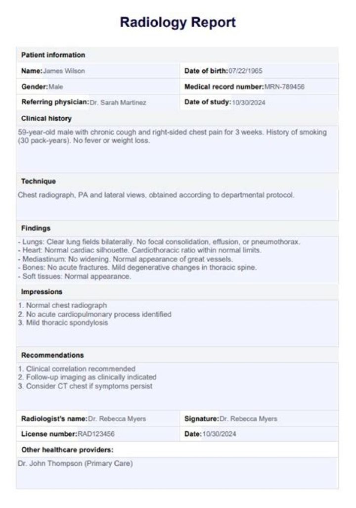
What does a radiologist report look like?
The radiology report is most often organized into 6 sections: type of exam, clinical information, comparison, technique, findings, impression. Let’s take these one at a time.
What are radiology reports?
The written radiology report is the most critical component of the service provided by a radiologist. It constitutes the formal documentation and communication of the results of a radiologic study or procedure.
Why is a radiology report important?
The written radiological report is the most important means of communication between the radiologist and referring medical doctor [1–3]. It is part of the patient’s permanent health record, and interprets the investigation in the clinical context.What is XRAY report?
Overview. An X-ray is a quick, painless test that produces images of the structures inside your body — particularly your bones. X-ray beams pass through your body, and they are absorbed in different amounts depending on the density of the material they pass through.
Can radiologist read MRI?
When you go in for a scan (whether it’s an MRI, an X-ray, computed tomography, or any other imaging modality), technologists obtain the images. When the images are complete, a radiologist examines, or “reads,” them, and writes a report indicating clinically significant details.
What tests are done in radiology?
- Computed tomography (CT) scan.
- Magnetic resonance imaging (MRI) scan.
- Breast MRI.
- X-rays and other radiographic tests.
- Mammography.
- Nuclear medicine scans (bone scans, PET scans, Thyroid scans, MUGA scans, gallium scans)
- Ultrasound.
What does the CXR normally look like in PE?
The classic radiographic findings of pulmonary infarction include a wedge-shaped, pleura-based triangular opacity with an apex pointing toward the hilus (Hampton hump) or decreased vascularity (Westermark sign). These findings are suggestive of pulmonary embolism but are infrequently observed.What does nonspecific mean in radiology report?
A “nonspecific symptom” is something that is reported by a patient but cannot be observed. Nonspecific symptoms may include chronic fatigue or pain that is not related to a known injury.
What are the 5 imaging techniques?Learn more about our five most common modalities for our various types of imaging tests: X-ray, CT, MRI, ultrasound, and PET.
Article first time published onWhat are modalities in radiology?
Medical imaging modalities, for example, includes magnetic resonance imaging (MRI), ultrasound, medical radiation, angiography and computed tomography (CT) scanners. In addition, to several scanning techniques to visualise the human body for diagnostic and treatment purposes.
What are XRAY services?
What Is an X-ray? An x-ray, which is short for x-radiation, is a type of imaging test that has been utilized for years. With this technology, doctors can see your bones, muscle and more without having to make an incision. This helps them diagnose, treat and monitor a number of different medical conditions.
How often do radiologists make mistakes?
Yes! It may shock you to learn that the error rate for radiologists is 4%. And on average there are 1 billion radiology exams each year. By this logic, that means there will be 40 million radiologist errors.
Do radiologists know results?
Unless the radiologist performs a history and physical examination, he will not know much about the patient. A lack of clinical context might cause a radiologist to misinform the patient.
Will a radiographer tell you if something is wrong?
The scan is performed by a tech. They pretty much know what they’re looking at but aren’t responsible for interpreting results and are not allowed to tell the patients what they see. The radiologist is a doctor, and he or she is the one that interprets the results and writes the report.
What does clinically indicated mean?
clinically indicated. The issue is rather: are findings observed on the. nuclear medicine procedure to suggest that an additional procedure is indi.
What does no clinical findings mean?
a : not relating to, involving, or concerned with the direct observation and treatment of living patients a nonclinical job nonclinical duties. b : not based on or characterized by observable and diagnosable symptoms of disease a nonclinical infection.
How do you read a MRI report?
- Start by checking the patient and image details.
- Look at all the available image planes.
- Compare the fat-sensitive with the water-sensitive images looking for abnormal signal.
- Correlate the MRI appearances with available previous imaging.
- Relate your findings to the clinical question.
How do you confirm PE?
- Blood tests. …
- Chest X-ray. …
- Ultrasound. …
- CT pulmonary angiography. …
- Ventilation-perfusion scan (V/Q scan) …
- Pulmonary angiogram. …
- MRI. …
- Medications.
What is a Hampton hump?
Hampton’s hump is a radiological sign consisting of a peripheral, wedge-shaped opacification adjacent to the pleural surface, which represents pulmonary infarction distal to a pulmonary embolus. 1. Owing to good pulmonary perfusion from collateral blood vessels, this sign is rarely seen in clinical practice.
What is the most common CXR finding in a patient with pulmonary embolism PE )?
The most common chest radiographic finding in patients with PE was atelectasis and/or parenchymal areas of increased opacity; however, the prevalence was not significantly different from that in patients without PE.
What are 3 types of medical imaging?
- X-rays.
- CT scans.
- Ultrasounds.
- Mammography.
- MRI.
What is the best imaging technique?
Diagnostic imaging techniques help narrow the causes of an injury or illness and ensure that the diagnosis is accurate. These techniques include x-rays, computed tomography (CT) scans, and magnetic resonance imaging (MRI).
How many types of radiography are there?
There are three types of diagnostic radiographs taken in today’s dental offices — periapical (also known as intraoral or wall-mounted), panoramic, and cephalometric. Periapical radiographs are probably the most familiar, with images of a few teeth at a time captured on small film cards inserted in the mouth.
What are the 3 earliest imaging modalities?
- The skeletal systems.
- The oral cavity (bone and teeth)
- Any ingested objects.
- The lungs.
- The breast (Mammography)
- The digestive system.
What is a Dicom modality?
DICOM is used worldwide to store, exchange, and transmit medical images. DICOM has been central to the development of modern radiological imaging: DICOM incorporates standards for imaging modalities such as radiography, ultrasonography, computed tomography (CT), magnetic resonance imaging (MRI), and radiation therapy.
What procedures does a radiologist perform?
Radiologists are medical doctors that specialize in diagnosing and treating injuries and diseases using medical imaging (radiology) procedures (exams/tests) such as X-rays, computed tomography (CT), magnetic resonance imaging (MRI), nuclear medicine, positron emission tomography (PET) and ultrasound.
What are the 5 most common errors in radiology?
- projection/technique errors (M)
- Exposure/processing errors.
- Film handling errors.
- Digital errors.
Why do radiologists disagree?
Radiologist can make mistakes due to the technical or physical limitations of the imaging modality. Staff shortages, staff inexperience and inadequate equipment are often the cause of errors.
Why do radiologists miss things?
Pathologists and clinical laboratory professionals who regularly analyze images will be interested in the findings of a research study designed to assess how the phenomenon called “inattentional blindness” among radiologists could cause them to possibly miss things hiding in plain sight.