How do you remember lumbar plexus
I: iliohypogastric nerve.I: ilioinguinal nerve.G: genitofemoral nerve.L: lateral femoral cutaneous nerve.O: obturator nerve.F: femoral nerve.L: lumbosacral trunk.
What is the lumbar plexus?
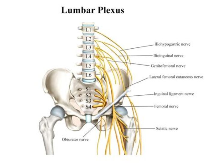
The lumbar plexus is an essential collection of nerves that arise from mostly the lumbar spinal cord. The term plexus refers to a “web” of nerves that is created just lateral to where T12-L5 exit the spinal cord via intervertebral foramina.
How do you remember your spinal levels?
You can use a meal-related mnemonic to remember them – imagine a crunchy breakfast at 7 am (7 cervical vertebrae), a tasty lunch at 12 noon (12 thoracic vertebrae), and a light dinner at 5 pm (5 lumbar vertebrae). Figure 6.13 The bones of the vertebral column.
What are the 3 branches of the lumbar plexus?
The lumbar plexus gives rise to several branches which supply various muscles and regions of the posterior abdominal wall and lower limb. These branches include the iliohypogastric, ilioinguinal, genitofemoral, lateral femoral cutaneous, femoral and obturator nerves.What is the lumbar plexus formed by?
Lumbar plexus is formed within the psoas major muscle from the ventral rami of the first four lumbar nerves and a contribution of the last thoracic nerve (T12). It forms the iliohypogastric, ilioinguinal, genitofemoral, lateral cutaneous nerve of thigh, obturator and femoral nerves.
What is the cauda?
Cauda is Latin for tail, and equina is Latin for horse (ie, the “horse’s tail”). The CE provides sensory innervation to the saddle area, motor innervation to the sphincters, and parasympathetic innervation to the bladder and lower bowel (ie, from the left splenic flexure to the rectum).
What is the location of the lumbar plexus?
Location. The lumbar plexus contains the ventral rami (front portions of spinal nerves) that emerge from between the five lumbar vertebrae (L1-L5). In addition, it’s joined by a portion of the lowest thoracic nerve, which emerges from the T12 vertebra just above the lumbar region.
What is anterior and posterior division of lumbar plexus?
The lumbar plexus is made up of anterior and posterior divisions. The anterior division gives rise to the ilioinguinal, iliohypogastric, and genitofemoral nerves, all of which are derived from L1–L2 and provide sensory innervation to the lower abdomen, upper proximal thigh, and lateral genitalia.Is the lumbar plexus sympathetic?
Four lumbar splanchnic nerves arise from sympathetic trunk to join the coeliac, inferior mesenteric and superior hypogastric plexuses. First lumbar splanchnic nerve arises from the first ganglion gives branches to coeliac, renal and inferior mesenteric plexuses.
What is cervical plexus?The cervical plexus is a network of nerve fibres that supplies innervation to some of the structures in the neck and trunk. It is located in the posterior triangle of the neck, halfway up the sternocleidomastoid muscle, and within the prevertebral layer of cervical fascia.
Article first time published onWhat does lumbar mean in medical terms?
Medical Definition of lumbar 1 : of, relating to, or constituting the loins or the vertebrae between the thoracic vertebrae and sacrum the lumbar region. 2 : of, relating to, or being the abdominal region lying on either side of the umbilical region and above the corresponding iliac region.
What is cord equina?
The cauda equina is the sack of nerve roots (nerves that leave the spinal cord between spaces in the bones of the spine to connect to other parts of the body) at the lower end of the spinal cord. These nerve roots provide the ability to move and feel sensation in the legs and the bladder.
What is phylum terminal?
The filum terminale (FT) is a fibrous band that extends from the conus medullaris to the periosteum of the coccyx, and its functions are to fixate, stabilize, and buffer the distal spinal cord from normal and abnormal cephalic and caudal traction.
What is saddle anesthesia?
Saddle anaesthesia refers to reduced sensation in the area that would be in contact with a saddle if sitting on one. This includes the perineum, buttocks, anus, groin and upper thighs. Saddle anaesthesia will make these areas feel numb and abnormal.
What is a lumbar sympathetic block?
A lumbar sympathetic block is an injection of medication that helps relieve lower back or leg pain (sciatica). It can be used to treat: Reflex sympathetic dystrophy. Complex regional pain syndrome. Herpes zoster infection (shingles) involving the legs.
What is a plexus?
A plexus is a bundle of intersecting nerves, blood vessels, or lymphatic vessels in the human body. These bundles typically originate from the same anatomical area and serve specific areas of the body. Bundles of nerves that form a plexus communicate information to your brain about pain, temperature, and pressure.
What forms cervical plexus?
The cervical plexus is formed from the anterior primary rami of C1–C4, deep to the sternocleidomastoid muscle and in front of the scalenus medius and levator scapulae muscles.
What are the three ascending pathways?
Ascending tracts are sensory pathways that begin at the spinal cord and stretch all the way up to the cerebral cortex. There are three types of ascending tracts, dorsal column-medial lemniscus system, spinothalamic (or anterolateral) system, and spinocerebellar system.
Is corticospinal tract ascending or descending?
The lateral corticospinal tract (LCST) is the largest descending motor pathway. It begins in the cerebral cortex, receiving a range of inputs from the primary motor cortex, premotor cortex and supplementary motor areas.
Is a medulla oblongata?
Medulla oblongataSection of the medulla oblongata at about the middle of the olivary bodyDetailsPart ofBrain stemIdentifiers
What does C1 and C2 innervate?
C1, C2, and C3 (the first three cervical nerves) help control the head and neck, including movements forward, backward, and to the sides.
How do spinal nerves exit the vertebral column?
The spinal nerves leave the vertebral column through the intervertebral foraminae. Some spinal nerves are intermingled in plexuses, from which the peripheral nerves are formed, each nerve containing fibers from several spinal cord segments.
What is innervated by C1?
Muscles innervated by this nerve are: … Geniohyoid muscle- through Hypoglossal nerve. Rectus capitis anterior muscle. Longus capitis muscle (partly)
What are the 4 nerve plexuses?
Of the four major nerve plexuses (cervical, brachial, lumbar, and sacral), only the brachial plexus and sacral plexus can be assessed satisfactorily in the EDX laboratory.
Which plexus Innervates the diaphragm?
Phrenic nerveThe phrenic nerve emerges from the cervical plexus, with the right brachial plexus shown here.DetailsFromC3–C5 of cervical plexusInnervatesDiaphragm
What muscles are innervated by c2 C3?
The muscular branches pass deeply from the plexus to supply the rhomboids, the serratus anterior, the sternocleidomastoid, the trapezius, levator scapulae, and the scalenus medius. There are also branches that supply the muscles of the suboccipital triangle.
Is lumbar spine the same as lumbosacral spine?
A lumbosacral spine x-ray is a picture of the small bones (vertebrae) in the lower part of the spine. This area includes the lumbar region and the sacrum, the area that connects the spine to the pelvis. This is the spine and the sacrum with the cervical (neck), thoracic (mid-back), and lumbar (lower back) vertebra.
What is it called when you have lower back pain?
Lower back pain, sometimes called lumbago, is not a specific disease diagnosis. It’s a symptom of several different types of medical problems. It usually results from a problem with one or more parts of the lower back, such as: ligaments and muscles.
What does an MRI of the lumbar spine show?
An MRI of the lumbar spine shows the bones, disks, spinal cord, and the spaces between the vertebral bones where nerves pass through.