How do you remove the baseline wander on an ECG
Baseline wander is a low-frequency noise of around 0.5 to 0.6 Hz. To remove it, a high-pass filter of cut-off frequency 0.5 to 0.6 Hz can be used. Powerline interference (50 or 60 Hz noise from mains supply) can be removed by using a notch filter of 50 or 60 Hz cut-off frequency.
What causes baseline drift in ECG?
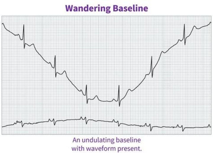
Baseline wander is a commonly seen noise in ECG recordings and can be caused by respiration, changes in electrode impedance, and motion. Baseline wander can mask important information from the ECG, and if it is not properly removed, crucial diagnostic information contained in the ECG will be lost or corrupted.
What are the principle causes of wandering baseline How do you remove the artifact?
The main cause of the BW in the ECG signal is movement and respiration of the patient [2]. One of the approaches to remove BW artefacts is the high-pass filtering of ECG signals [2].
What is baseline wandering in ECG?
Baseline wander is a low frequency artifact in the ECG that arises from breathing, electrically charged electrodes, or subject movement and can hinder the detection of these ST changes because of the varying electrical isoline (Figure 1(a)).How do you remove motion artifacts from ECG?
An existing method to remove the motion artefact is to employ an accelerometer for measuring the body movement at the same time of ECG detection [5]. However, for non-contact electrode structure of ECG detection, an accelerometer directly attached to the human body is unacceptable.
What causes an interrupted baseline?
It can be caused by patient movement, including breathing. I have also noticed that stopping or accelerating the ambulance can cause wandering baseline. Some references suggest that wandering baseline can be caused by loose or dry electrodes.
What does baseline drift mean?
Baseline drift is the low-frequency signal variation that occurs in the baseline due to column stationary phase bleed, background ionization, and low-frequency variations in the detector and/or instrument-controlled parameters (such as temperature or flow).
How do you find the baseline of an ECG?
If you follow the QRS complex on your ECG, you will see that they are usually sharp-pointed. If you go down with the Q wave, up with the R wave, down the S wave and follow the S wave back to the baseline, it will usually pass the baseline. The moment that line goes horizontal, that is where your J point is.What is base line wandering how can it be prevented in line coders?
Line coding technique to eliminate baseline wandering In the first half, voltage is at one level and in the second half it is at the other level. Transition at the center of bit period helps in synchronization. Differential Manchester encoding combines RZ and NRZ-I. There is transition at the center of the bit period.
What are baseline ECG abnormalities?Common baseline abnormalities included sinus bradycardia, R wave transition abnormalities, right axis deviation, non-specific T wave changes and atrial premature complexes.
Article first time published onWhen a PVC appears on the ECG which wave is missing?
The PVC appears on every second beat. The EKG rhythm will appear irregular. The P wave is absent and the PR interval is not measurable. The QRS complex will typically be wide (> 0.10 sec) and bizarre in appearance.
How can you reduce artifacts?
- Minimize the degree of motion. a. The importance of simple instruction/education of the patient to hold still while the scanner is making noise should not be underestimated. …
- Suppress signal from moving tissues. a. …
- Adjust imaging sequences and parameters. a. …
- Detect and compensate for motion.
What is the effect of motion artifacts in ECG recordings?
Electrocardiographic artifacts are defined as electrocardiographic alterations, not related to cardiac electrical activity. As a result of artifacts, the components of the electrocardiogram (ECG) such as the baseline and waves can be distorted. Motion artifacts are due to shaking with rhythmic movement.
How does movement affect an ECG?
It is important to be relaxed and warm during an ECG recording because any movement, including shivering, can alter the results. Sometimes this test is done while you are exercising or under light stress to look for changes in the heart. This type of ECG is often called a stress test.
How do I fix my baseline drift?
- Use a heat exchanger before the detector to control the temperature of the column and mobile process.
- Use HPLC-grade solvents, high-purity salts, and additives. …
- Flush cell with methanol or other potent solvents. …
- Unplug or replace the row. …
- Correct rate of composition/flow.
What is the purpose of the patient diary in Holter monitoring?
One of the most critical parts of the Holter monitor test is the patient diary. Patients are asked to keep a diary recording the timing and type of symptoms they have. These are then correlated with the tracings to see if symptoms and tracing abnormalities are related.
What causes electrical interference on ECG?
Another disturbance or annoyance in terms of rhythm detection, emanating directly from the surrounding environment , is electrical interference. The ECG machine is designed to pick up electrical activity within the heart but it will pick up electrical activity from nearby machinery, such as: Pumps.
Which arrhythmia is viewed as a common disruption in rhythm?
Atrial fibrillation (AF) is a common heart rhythm disorder caused by degeneration of the electrical impulses in the upper cardiac chambers (atria) resulting in a change from an organized heart rhythm to a rapid, chaotic rhythm.
Which scheme avoids the drawback of baseline wandering?
The Manchester scheme overcomes several problems associated with NRZ-L, and differential Manchester overcomes several problems associated with NRZ-I as there is no baseline wandering and no DC component because each bit has a positive and negative voltage contribution.
What is baseline wandering DC components and frequency domain?
1) Baseline wandering: The receiver averages the signal power (Baseline), and uses it to decode the received signal bit value. 2) DC components: Constant level for long period of time creates very low frequency components in the frequency spectrum, that might not pass through some medium (e.g., TP of 200Hz→ 3000Hz).
Which line coding is best?
Differential encoding: In many cases if the leads from a device are accidentally inverted and connected all the ones and zeros may be inverted; for this differential encoding is the best solution. Because, it allows the polarity of a signal to be inverted with out affecting the data detection.
What is a deflection from the baseline?
By convention, the first upward deflection from the baseline is termed the P wave, and it reflects atrial depolarization. The P wave should not exceed 2.5 mm in height nor 0.11 second in width (i.e., less than three small boxes high and wide). Ventricular depolarization is represented by the QRS complex.
When should you get a baseline EKG?
You should probably have an ECG if you have risk factors for an enlarged heart such as high blood pressure or symptoms of heart disease, such as chest pain, shortness of breath, an irregular heartbeat or heavy heartbeats.
How often are EKGS wrong?
The study of 500 patients found a false positive reading between 77 and 82 percent in patients screened by electrocardiogram, and a false negative reading between 6 percent to 7 percent in the same patient population.
Can anxiety cause an abnormal EKG?
Abnormal ECG Findings Caused by Anxiety Whether it is due to short-term test nervousness or a chronic condition, anxiety may be associated with certain ECG abnormalities, including T-wave inversion.
How does PVC look on ECG?
This is visible on the ECG as an inverted P wave (“retrograde P wave“), usually occurring after the QRS complex. PVCs are said to be “frequent” if there are more than 5 PVCs per minute on the routine ECG, or more than 10-30 per hour during ambulatory monitoring.
What does the small block on ECG paper represent when measuring the block horizontally?
ECG paper is a grid where time is measured along the horizontal axis. Each small square is 1 mm in length and represents 0.04 seconds. Each larger square is 5 mm in length and represents 0.2 seconds.
When PVCs appear in a pattern where every third beat is a PVC The pattern is called?
Sometimes, an extra heartbeat that starts in the lower ventricle disturbs your regular heart rhythm. It’s known as a premature ventricular contraction (PVC, also premature ventricular complex). When PVC happens in a pattern of three beats, doctors call it trigeminy.
How can an EEG EKG artifact be reduced?
- 3.1. Regression Methods. The traditional method for removing artifacts from EEG is the regression methods [37]. …
- 3.2. Wavelet Transform. …
- 3.3. BSS. …
- 3.4. Empirical Mode Decomposition. …
- 3.5. Filtering Methods. …
- 3.6. Sparse Decomposition Methods.
What are motion artifacts?
When a patient moves, it can cause distortion on the image, which is referred to as a motion artifact. Motion artifacts may appear as a blurring of contrast or edges, replication of part or all of a structure, signal loss or undesired strong signals.
What causes muscle artifacts?
Forehead, jaw, and eyelid muscle movements can cause artifacts by moving the electrodes. Movements in the surroundings produce disturbances by altering the ambient electrical fields. Moreover, the tongue and eyes have their own dipole electric charge.