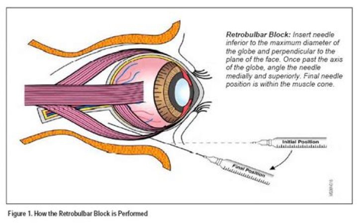
How is Retrobulbar block done
In this technique, local anesthetic is injected into the retrobulbar space, the area located behind the globe of the eye. This injection provides akinesia of the extraocular muscles by blocking cranial nerves II, III, and VI, which prevents movement of the globe.
How do they do an eye block?
What is a retrobulbar block? Retrobulbar block is an anesthetic procedure used for eye surgeries. Usually, lidocaine with epinephrine is injected into the retrobulbar space, which is the area located behind the eyeball (globe). It numbs the muscles around the eye by blocking cranial nerves II, III, and VI.
Does a Retrobulbar block hurt?
Most commonly, patients will report discomfort during the performance of the block, such as the sensation of the needle during insertion and/or pressure behind the eye during injection. In recent years, peribulbar block has become increasingly used because of its lower incidence of complications.
What nerves are blocked in a Retrobulbar block?
Retrobulbar block involves depositing local anesthetic inside the muscle cone. It aims to block the ciliary nerves, the ciliary ganglion, and cranial nerves III, IV, and VI.What is the meaning of retrobulbar?
Medical Definition of retrobulbar : situated, occurring, or administered behind the eyeball a retrobulbar injection.
Is Retrobulbar injection safe?
Retrobulbar anesthesia, competently administered, is a safe procedure. In 13 000 patients in whom a curved needle technique was used, the only serious complication was a single case of postoperative ischemic neuropathy [514].
What is retrobulbar haemorrhage?
Retrobulbar hemorrhage (RBH) is a rapidly progressive, sight-threatening emergency that results in an accumulation of blood in the retrobulbar space.
How do they deaden the eye for cataract surgery?
The eye drops act as an anesthetic. As you blink, the drops spread over your eye, numbing the surface. This allows you to feel no pain or discomfort during the surgery. When the eye is completely numb, an instrument will be used to hold your eye open while the procedure is completed.What is Peribulbar and Retrobulbar?
Peribulbar anaesthesia is performed by injecting the anaesthetic drug in the orbit around the equator of the eye ball (globe). Retrobulbar anaesthesia is performed by injecting the anaesthetic drug in the orbit further back behind the eye ball, which is near the nerves that control eye movement and sensation.
What is sub Tenon anesthesia?The sub-Tenon´s space is a virtual space between the capsule and the sclera. The instillation of local anesthetic into this space produces analgesia and akinesia by diffusing posteriorly into the retro-orbital space to block the traversing sensory and motor nerves.
Article first time published onWhat is Intracameral?
An intracameral injection is usually of an antibiotic into the anterior chamber of the eyeball to prevent endophthalmitis caused by an infection of the eye that can occur after cataract surgery. The Food and Drug Administration (FDA) has not approved antibiotics for this use and it is considered ‘off-label’.
How do you give a retrobulbar injection?
Retrobulbar injection: the needle is passed through the junction of the middle and outer third of the inferior orbital rim, then straight back below the eye for 15 mm. The needle should be parallel to the floor of the orbit and angled down.
How is anesthesia used for eye surgery?
Local anaesthesia for an eye operation For eye surgery, it can be given as eye drops and/or injections. After you have the local anaesthetic you will still be awake and aware of what is happening to you. The aim is that you feel no pain during the operation.
How long does a nerve block last after cataract surgery?
Depending on the type of anesthetic, a block may last over four hours with a mixture of lidocaine 1% and bupivicaine 0.375%. Addition of hyaluronidase to the anesthetic mix improves penetration of anethesia (Anesth Analg 2000 Oct;91(4):934-7.)
What is the meaning of bulbar?
Bulbar: Pertaining to a bulb, in medicine any rounded mass of tissue (that is shaped somewhat like a crocus or tulip bulb). For example, the bulbar conjunctiva is that part of the conjunctiva, a clear membrane of the eye, which covers the outer rounded surface of the eye. Bulbar can also apply to a rounded enlargement.
What causes Retrobulbar hemorrhage?
What are the common causes and possible consequences? A retrobulbar hemorrhage is bleeding that occurs posterior to the orbital septum or globe in sufficient quantity to exert pressure on the globe. It may result from trauma, orbital surgery, and especially the removal of fat during blepharoplasty.
How do you treat a retrobulbar hematoma?
The majority of retrobulbar hemorrhages can be managed conservatively with digital ocular massage or intravenous acetazolamide or mannitol. However, further surgical intervention is indicated when vision is at risk.
How do you do a lateral Canthotomy?
Use iris scissors to cut from the lateral canthus to the rim of the orbit, about 1 to 2 cm (canthotomy). Cut the inferior and sometimes both crus of the lateral canthal ligament (cantholysis). Most experts recommend starting with the inferior crus. Lift the lateral portion of lower eyelid.
How is retrobulbar optic neuritis diagnosed?
- Blurred or dimmed vision.
- A blind spot at or near the center of vision.
- Color “wash-out” so that colors are less rich.
- Pain with eye movement.
- Tenderness of the eye to touch or pressure.
- Complete blindness in the affected eye.
What are Retrobulbar headaches?
Retrobulbar neuritis is a form of optic neuritis in which the optic nerve, which is at the back of the eye, becomes inflamed. The inflamed area is between the back of the eye and the brain. The optic nerve contains fibers that carry visual information from the nerve cells in the retina to the nerve cells in the brain.
What causes retrobulbar optic neuritis?
Retrobulbar optic neuritis (RON) is mainly caused by multiple sclerosis, a common demyelinating disease. The cardinal signs of RON are the loss including visual acuity or/and contrast sensitivity, periocular pain induced with ocular movements, RAPD and CVD.
What is the CPT code for infraorbital nerve block?
There is also a code for trigeminal nerve block for dental pain (CPT code 64400, $130 on the Medicare Physician Fee Schedule). This includes blocks for the infraorbital and inferior alveolar nerves. Want to read more about nerve block reimbursement?
When do you give an infraorbital nerve block?
Nerve blocks are useful for achieving anesthesia to a regional area of the body. Regional nerve blocks offer many advantages over local tissue infiltration. They are useful when local infiltration may not be possible or could result in tissue damage or distortion.
How do you give an ASA nerve block?
Approach: While retracting the lip, insert the needle into the intersection of the mucobuccal fold and the apex/center of the canine at a 45-degree angle, advancing the needle approximately 1-1.5 cm. Aspirate. Slowly inject 2 mL of local anesthetic and massage for 10-20 seconds.
What is the difference between Peribulbar and retrobulbar block?
Peribulbar anaesthesia is performed by injecting the anaesthetic drug in the orbit around the equator of the eye ball (globe). Retrobulbar anaesthesia is performed by injecting the anaesthetic drug in the orbit further back behind the eye ball, which is near the nerves that control eye movement and sensation.
What is a Honan balloon?
The Honan balloon is well known in the field of ophthalmic anesthesia. It is used as a compression device after injection of local anesthesia to help in the diffusion of the anesthesia and to reduce intraocular pressure (IOP).
How long does a cataract operation take?
Cataract surgery takes 10 to 20 minutes to complete, depending on the severity of the condition. You should also plan to spend up to 30 minutes following the surgery to recover from the effects of the sedative.
Are you put to sleep when you have cataract surgery?
Typically, patients are awake during cataract surgery. This eliminates risks associated with general anesthesia (where you are “put to sleep”) and enables Our Doctors to communicate with you during your procedure. You will be given an oral medication prior to the procedure to help you relax during your surgery.
Are you sitting or lying down during cataract surgery?
The standard position for patients having cataract surgery is the supine position: the patients lie flat on their backs to face the operating microscope overhead.
How long does Subtenon block last?
This combination will provide approximately 60–90 minutes of surgical anesthesia and 4–6 hours of postoperative analgesia. Other options include 2% lignocaine/150 iu hyauronidase, which will provide approximately 45 minutes of surgical anesthesia and is useful for short cases and faster cataract surgeons.
What is Tenon's capsule?
Tenon’s capsule is a dense, elastic, and vascular connective tissue layer that surrounds the globe except over the cornea, and invests the anterior portions of the extraocular muscles.