How is tarsal coalition diagnosed
Computerized tomography scan (CT or CAT scan): Considered the gold standard for diagnosing tarsal coalitions, a CT scan is a diagnostic imaging procedure that uses a combination of x-rays and computer technology to produce cross-sectional horizontal and vertical images (called “slices”) of the body.
How do I know if I have a tarsal coalition?
- Pain and stiffness in the foot and ankle, particularly when your child is walking or standing.
- A rigid flat foot, which may make walking more difficult.
- Walking with a limp.
- Muscle spasms in the leg, which can cause the foot to turn inward.
- Frequent sprained ankles.
How do you rule out the tarsal coalition?
- X-rays. These tests provide clear images of bone. …
- Computed tomography (CT) scans. The images produced with computed tomography provide greater detail of the bones.
Does tarsal coalition show up on xray?
Tarsal coalitions may be osseous, cartilaginous, or fibrous. Calcaneonavicular coalitions are readily detected on oblique radiographs. … Moreover, CT or MR imaging may be required to confirm the diagnosis of talocalcaneal coalition when radiographic findings are equivocal.What does tarsal coalition feel like?
pain, typically on the outside and top of the foot (though some children have no pain) flat feet or a flat foot (though not all children with flat feet have a tarsal coalition) rigidity and stiffness in the affected foot. muscle spasms.
What is suspected tarsal coalition?
A tarsal coalition is an abnormal connection that develops between two bones in the back of the foot (the tarsal bones). This abnormal connection, which can be composed of bone, cartilage or fibrous tissue, may lead to limited motion and pain in one or both feet.
Is tarsal coalition considered a disability?
Tarsal coalitions may cause altered foot biomechanics leading to patient disability from osteoarthritis and other sequelae. While some types of coalition are common, isolated talonavicular coalitions are relatively rare.
What is tarsal coalition surgery?
Removal of the Tarsal Coalition The surgery involves simply removing the abnormal tissue to allow motion of the back part of the foot. A soft tissue spacer, such as fat or tendon, is placed at removed coalition site to limit bone re-growth. This surgery preserves the rearfoot joints.Can you play sports with tarsal coalition?
Your child can benefit from the exercise and team building that results from participation in organized sports. Conditions like tarsal coalition in athletes (fusing of tarsal bones), though, can put a damper on their enthusiasm.
Is tarsal coalition congenital?Congenital tarsal coalition is a diagnosis that is often overlooked in young patients who first present with foot and ankle pain. Calcaneonavicular and talocalcaneal coalitions are encountered most frequently; fusion at other sites is much less common. Tarsal coalitions may be osseous, cartilaginous, or fibrous.
Article first time published onHow can I help the tarsal coalition pain?
- Nonsteroidal anti-inflammatory drugs (NSAIDs) such as ibuprofen to reduce pain and inflammation.
- Physical therapy, including massage, range-of-motion exercises and ultrasound therapy.
- Steroid injection(s) into the affected joint to reduce pain and inflammation.
How long does it take to recover from tarsal coalition?
This procedure may or may not require using a cast after surgery. The recovery time may take 6-12 months.
Does tarsal coalition cause plantar fasciitis?
There are many possible causes for foot pain, from plantar fasciitis to fractures, foot neuromas and bone spurs. One such cause is a tarsal coalition. Dr. Stuart Katchis, NY-based orthopedic surgeon specializing in the foot and ankle, has treated many young patients with tarsal coalitions.
Is tarsal carpal coalition syndrome curable?
Symptoms of TCC may include: stiffness and progressive immobility of the hands and feet and short stature . TCC is caused by mutations in the NOG gene , and it is inherited in an autosomal dominant manner. Although there is no specific treatment or cure for TCC, there may be ways to manage the symptoms.
Where is Tarsus in foot?
In the human body, the tarsus is a cluster of seven articulating bones in each foot situated between the lower end of the tibia and the fibula of the lower leg and the metatarsus. It is made up of the midfoot (cuboid, medial, intermediate, and lateral cuneiform, and navicular) and hindfoot (talus and calcaneus).
How is Calcaneonavicular coalition treated?
Summary. Calcaneonavicular coalition is a common source of pain and more or less severe flat and stiff foot in children. Classically, treatment consists in resecting the coalition using a dorsolateral approach. Good quality resection and interposition can prevent recurrence.
Why does my tarsal coalition hurt?
The tarsal coalition may be associated with a stiff flatfoot. Pain may come not only from the coalition itself, but from the peroneal tendons (which sit around the outside of the ankle). These tendons can shorten and go into spasm. Ligament strain and joint irritation may also generate pain.
Does tarsal coalition always affect feet?
It affects both feet about 50% of the time and is not always symptomatic. The cause of tarsal coalition is unknown. with other conditions. in about 1% of the population.
Can you have tarsal tunnel syndrome in both feet?
If you answer yes to either or both of these questions, you may have a condition known as tarsal tunnel syndrome. This syndrome can occur in one foot or both feet and is similar to the common carpel tunnel syndrome in the hand.
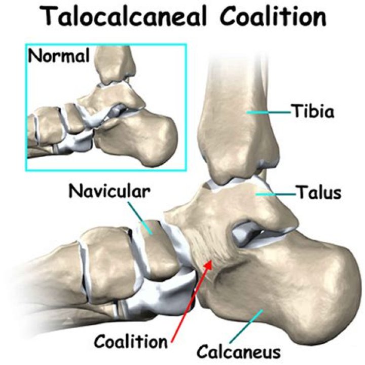
How is Talocalcaneal coalition treated?
Conclusions: A symptomatic talocalcaneal coalition can be treated with excision and fat graft interposition, and achieve good to excellent results in 85% of patients. Patients should be counseled that a subset may require further surgery to correct malalignment.
What are the 7 tarsal bones?
The tarsal bones are 7 in number. They are named the calcaneus, talus, cuboid, navicular, and the medial, middle, and lateral cuneiforms.
What is cuboid navicular coalition?
Cuboid-navicular coalition, a type of tarsal coalition, is extremely rare with less than 10 reported cases to date. Most prevailing theories reported have described this specific type of coalition as asymptomatic except at specific moments of stress and exercise.
How common is accessory navicular syndrome?
An accessory navicular is an extra bone that is on the inner center arch of the foot. Up to 2.5 percent of individuals are born with the accessory navicular.
What is subtalar coalition?
Talocalcaneal coalition or peroneal spastic foot or subtalar coalition is an anomalous connection between the talus and the calcaneum that can present with painful and rigid flat-foot in older children and adolescents. The talocalcaneal coalition is part of a spectrum of tarsal coalitions that causes rigid flat foot.
When can I walk after foot surgery?
Since virtually all foot and ankle operations require rest and elevation of the operated foot for at least 2 weeks following surgery, it is rare that a patient will be allowed to return to work before 2 weeks following surgery.
Is tarsal the ankle?
The tarsal tunnel is located on the inside of the ankle, and is formed by the ankle bones and the band of ligaments that stretches across the foot. Many of the blood vessels, nerves and tendons that provide movement and flexibility to the foot travel through the tarsal tunnel.
What is tarsus bone?
tarsal, any of several short, angular bones that in humans make up the ankle and that—in animals that walk on their toes (e.g., dogs, cats) or on hoofs—are contained in the hock, lifted off the ground. The tarsals correspond to the carpal bones of the upper limb.
What is subtalar fusion?
Subtalar fusion is the best procedure to correct long-term pain caused by injury or arthritis. The surgery involves the process of fusing the subtalar joint to the adjacent ankle joint.
What is talar beaking?
The term ‘talar beak’ refers to a flaring of the superior aspect of the talar head, seen on lateral radiographs. 2. This is an indirect sign of talocalcaneal coalition and thought to form as a consequence of impaired subtalar joint motion, which results in the navicular overriding the talus.
What is ankle fusion surgery?
Ankle fusion is a type of surgery to fuse the bones of your ankle into one piece. It’s also known as ankle arthrodesis. The surgery is usually done to treat arthritis in the ankle. The ankle joint is also called the tibiotalar joint. It’s where the shinbone (tibia) rests on top of a bone of the foot called the talus.
What gene causes tarsal coalition?
Tarsal-carpal coalition syndrome is caused by mutations in the NOG gene, which provides instructions for making a protein called noggin. This protein plays an important role in proper bone and joint development by blocking (inhibiting) signals that stimulate bone formation.