How much is Gerd Binnig worth
Gerd Binnig, (born July 20, 1947, Frankfurt am Main, W. Ger.), German-born physicist who shared with Heinrich Rohrer (q.v.) half of the 1986 Nobel Prize for Physics for their invention of the scanning tunneling microscope.
What did Gerd Binnig do?
Gerd Binnig, (born July 20, 1947, Frankfurt am Main, W. Ger.), German-born physicist who shared with Heinrich Rohrer (q.v.) half of the 1986 Nobel Prize for Physics for their invention of the scanning tunneling microscope.
What did Gerd Binnig and Heinrich Rohrer do?
Swiss physicist Heinrich Rohrer co-invented the scanning tunneling microscope (STM), a non-optical instrument that allows the observation of individual atoms in three dimensions, with Gerd Binnig. The achievement garnered the pair half of the Nobel Prize in Physics in 1986.
Who inspired Gerd Binnig?
He began playing violin at age 15 and although he did not develop a great talent, he enjoyed playing in the school orchestra nonetheless. Influenced by his older brother’s immersion in such bands as the Rolling Stones and the Beatles, Binnig played in a rock band with friends and even wrote some of his own music.What did Rohrer and Binnig call their microscope?
With the introduction of these changes, Binnig and Rohrer turned the instrument into a microscope and called it Scanning Tunneling Microscope. It is not clear how exactly they came to the idea of transforming their tunneling testing instrument into an STM, displaying images of surfaces on the atomic scale.
Who invented tunneling microscope?
In 1981, two IBM researchers, Gerd Binnig and Heinrich Rohrer, broke new ground in the science of the very, very small with their invention of the scanning tunneling microscope (STM).
How much does a scanning tunneling microscope cost?
Low cost and relatively low quality STMs start at approximately $8,000 but some people have actually built their own amateur STMs for much less than that amount. However, professional quality STMs can range anywhere from $30,000 to $150,000 depending on the manufacturer and the extra parts included.
Who is Gerd Binnig and Heinrich Rohrer?
Gerd Binnig and Heinrich Rohrer invented the Scanning Tunneling Microscope in 1981 working at IBM Zurich. Binnig also invented the Atomic Force Microscope with Calvin Quate in 1986 while spending a year at Stanford University. Binnig and Rohrer received the Nobel Prize for physics in 1986.IS STM or SEM better?
The scanning tunneling microscope (STM) differs significantly from the SEM. It is capable of imaging objects at ten times the lateral resolution, to 0.1 nanometer. … An STM at the London Centre for Nanotechnology. The central concept in the STM is that of a small conducting tip brought near to the sample.
Who won Nobel Prize in physics from his invention of phase contrast microscope?The Nobel Prize in Physics 1953 was awarded to Frits Zernike “for his demonstration of the phase contrast method, especially for his invention of the phase contrast microscope.”
Article first time published onWhat year was the scanning tunneling microscope invented?
September 1981: Invention of the scanning tunneling microscope.
Who invented tem?
Ernst Ruska at the University of Berlin, along with Max Knoll, combined these characteristics and built the first transmission electron microscope (TEM) in 1931, for which Ruska was awarded the Nobel Prize for Physics in 1986.
Who discovered STM in 1980?
scanning tunneling microscope (STM), device for studying and imaging individual atoms on the surfaces of materials. The instrument was invented in the early 1980s by Gerd Binnig and Heinrich Rohrer, who were awarded the 1986 Nobel prize in physics for their work.
Who is the father of microscopy?
Antoni van Leeuwenhoek (1632-1723): father of microscopy.
What is the cost of a scanning electron microscope?
The cost of a scanning electron microscope (SEM) can range from $80,000 to $2,000,000. The cost of a transmission electron microscope (TEM) can range from $300,000 to $10,000,000. The cost of a focused ion beam electron microscope (FIB) can range from $500,000 to $4,000,000.
Can you see atoms using a scanning tunneling microscope?
The wavelength of visible light is more than 1000 times bigger than an atom, so light cannot be used to see an atom. Scanning Tunneling Microscopes work by moving a probe tip over a surface we want to image. The probe tip is an extremely sharp – just one or two atoms at its point.
How much does an atomic force microscope cost?
Before they developed nGauge, traditional Atomic Force Microscopes (AFMs) cost anywhere between $100,000 to $500,000. AFMs are the workhorse of nanotechnology, and typically every university will have a few of these niche instruments on campus.
What does scanning tunneling microscope tell you?
Scanning Tunneling Microscopy allows researchers to map a conductive sample’s surface atom by atom with ultra-high resolution, without the use of electron beams or light, and has revealed insights into matter at the atomic level for nearly forty years.
Can we see atoms?
Atoms are really small. So small, in fact, that it’s impossible to see one with the naked eye, even with the most powerful of microscopes. … Now, a photograph shows a single atom floating in an electric field, and it’s large enough to see without any kind of microscope. 🔬 Science is badass.
Who won a Nobel Prize in 1986 for the scanning tunneling microscope?
The Nobel Prize in Physics 1986 was divided, one half awarded to Ernst Ruska “for his fundamental work in electron optics, and for the design of the first electron microscope”, the other half jointly to Gerd Binnig and Heinrich Rohrer “for their design of the scanning tunneling microscope.”
What can you see with a TEM microscope?
The transmission electron microscope is used to view thin specimens (tissue sections, molecules, etc) through which electrons can pass generating a projection image. The TEM is analogous in many ways to the conventional (compound) light microscope.
What is the difference between a SEM and TEM microscope?
The main difference between SEM and TEM is that SEM creates an image by detecting reflected or knocked-off electrons, while TEM uses transmitted electrons (electrons that are passing through the sample) to create an image.
Is TEM destructive?
However, a major limitation with TEM is the time-consuming, destructive sample preparation necessary for generating electron transparent specimens. … Scanning electron microscopy (SEM) has the significant advantage over TEM of being non- destructive and can rapidly image large areas.
What did Heinrich Rohrer do?
Heinrich Rohrer, who shared the 1986 Nobel Prize in Physics for inventing a microscope that made it possible to see individual atoms and move them around, an achievement that led to vastly faster computing and greatly advanced molecular biology, died on Thursday night or early Friday morning in Wollerau, Switzerland.
What did Heinrich Rohrer invent?
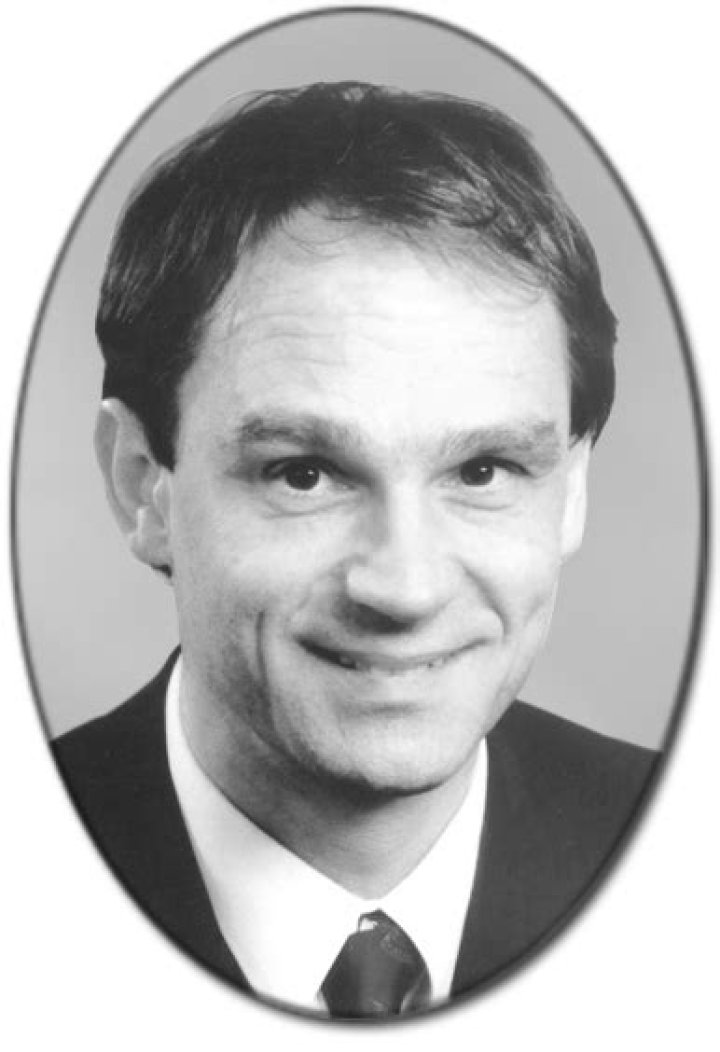
Heinrich Rohrer, (born June 6, 1933, Buchs, Switzerland—died May 16, 2013, Wollerau), Swiss physicist who, with Gerd Binnig, received half of the 1986 Nobel Prize for Physics for their joint invention of the scanning tunneling microscope.
What type of microscopy is phase contrast microscopy?
Phase-contrast microscopy (PCM) is an optical microscopy technique that converts phase shifts in light passing through a transparent specimen to brightness changes in the image. Phase shifts themselves are invisible, but become visible when shown as brightness variations.
When would you use the phase-contrast microscope?
- Living cells (usually in culture)
- Microorganisms.
- Thin tissue slices.
- Fibres.
- Subcellular particles, including organelles.
What type of microscope is usually used in school?
The most common types of microscopes used in teaching are monocular light microscopes (80%), followed by binocular optical microscopes (16%), digital microscopes (3%), and stereomicroscopes (1%). A total of 43% of teachers perform microscopy using the demonstration method, and 37% of teachers use practical work.
What type of samples can be imaged using STM?
Because STM is based on measuring the current between the tip and the sample, STM can only analyze conductor and semiconductor samples. Also, because most of the current is generated between the most outward atoms of the tip and the surface, atomic images can be generated only for atomically flat samples.
What is the difference between STM and AFM?
AFM refers to Atomic Force Microscope and STM refers to Scanning Tunneling Microscope. Unlike the STM, the AFM does not measure the tunneling current but only measures the small force between the surface and the tip. … It has also been seen that the AFM resolution is better than the STM.
Which of those constituents belong to a tem?
A TEM is composed of several components, which include a vacuum system in which the electrons travel, an electron emission source for generation of the electron stream, a series of electromagnetic lenses, as well as electrostatic plates.