Is blood white or black on CT
Look for any evidence of bleeding throughout all slices of the head CT. Blood will appear bright white and is typically in the range of 50-100 Houndsfield units.
Is blood dark on CT?
Blood products are absorbed with time (after two weeks) and change to clear liquid. It now appears as dark or hypodense in a pre-contrast CT scan. subacute subdural: In the subacute phase (between 3-14 days) it is isodense with brain and can easily be missed.
What color is blood on non contrast CT?
The CT scan demonstrates a subacute right frontoparietal subdural hematoma (red arrows). On noncontrast CT, acute blood is hyperdense and old blood is hypodense, so as the blood ages there will be a period of time when the blood is isodense to the brain parenchyma, as in this case.
Why does blood show up white on CT?
Acute haemorrhage absorbs X-rays and appears hyperdense (white) on CT scans. As the clot retracts it becomes more hyperdense over the first few hours up to 7 days; then isodense with brain over the following 1-4 weeks and finally hypodense compared with brain over the subsequent 4-6 weeks.What shows up black on CT scan?
PHYSICS OF CT The density of the tissue is in proportion to the attenuation of the x-rays which pass through. Tissues like air and water have little attenuation and are displayed as low densities (dark), whereas bone has high attenuation and is displayed as high density (bright) on CT.
What color is air on CT scan?
Bones appear white on the x-ray. Soft tissue, such as the heart or liver, shows up in shades of gray. Air appears black. With CT scanning, several x-ray beams and electronic x-ray detectors rotate around you.
What color is blood on CT?
Step 1: Blood Look for any evidence of bleeding throughout all slices of the head CT. Blood will appear bright white and is typically in the range of 50-100 Houndsfield units. Basic categories of blood in the brain are epidural, subdural, intraparenchymal/intracerebral, intraventricular, and subarachnoid.
Which side is which on CT?
When you look at a CT scan, it is like looking in a mirror. The right side of your body will be on the left side of the film and the left side of your body will be on right.Is blood Hyperdense on CT?
Hyperdensity at CT was due to the high hemoglobin content of retracted clot or sedimented blood. The various patterns seen can be related to sequential changes occurring in blood following hemorrhage. Relative hyperdensity and its variations seen on precontrast scans are useful diagnostic signs of recent hemorrhage.
What is white on CT head?bone to the detectors, resulting in its ‘white’ appearance on CT. ‡ White matter is less cellular, contains myelinated. axons (fat), and has a higher water content than. gray matter, resulting in slightly lower.
Article first time published onWhat does an active bleed look like on CT?
CONCLUSION. Active hemorrhage in patients after blunt abdominal trauma is most frequently visible as a jet of extravasated contrast agent on multidetector CT. When extravasation is detected, immediate surgical or angiographic therapy is required.
How can you tell the difference between a contrast and non contrast CT?
CT scans may be done with or without “contrast.” Contrast refers to a substance taken by mouth or injected into an intravenous (IV) line that causes the particular organ or tissue under study to be seen more clearly. Contrast examinations may require you to fast for a certain period of time before the procedure.
Can a CT detect bleeding?
CT angiography is an accurate examination for identifying the source of acute GI bleeding. A meta-analysis of data from 672 patients with moderate to severe UGIB and/or LGIB revealed an overall sensitivity of 85% and a specificity of 92% for detection of the bleeding site.
What shows up as white on a CT scan?
Bones appear white on the x-ray. Soft tissue, such as the heart or liver, shows up in shades of gray. Air appears black. With CT scanning, several x-ray beams and electronic x-ray detectors rotate around you.
What does a spot on a CT scan mean?
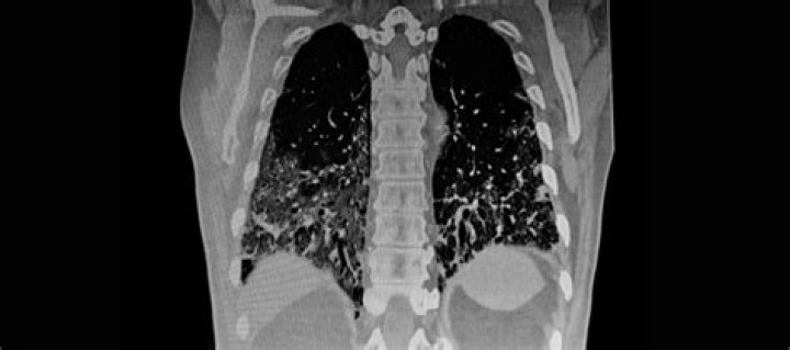
A spot on the lungs usually refers to a pulmonary nodule. This is a small, round growth on the lungs that shows up as a white spot on image scans. Typically, these nodules are smaller than three 3 centimeters (cm) in diameter. If your doctor sees a pulmonary nodule on a chest X-ray or CT scan, don’t panic.
What can a CT scan with contrast show?
A special dye called contrast material is needed for some CT scans to help highlight the areas of your body being examined. The contrast material blocks X-rays and appears white on images, which can help emphasize blood vessels, intestines or other structures.
What does black mean in CT?
On CT scans, the density of the image (with darker tissues being less dense) is attributed to x-ray attenuation. At the extremes of the CT density spectrum are air (black) and bone (white).
Why are CT scans black and white?
A CT scan uses a pencil-thin beam to create a series of pictures taken from different angles. The information from each angle is fed into a computer, which then creates a black and white picture that shows a slice of a certain area of the body – much like looking at a single slice from a loaf of bread.
What color is tumor on CT?
Air is black on CT (Figure 2a) and on all MR pulse sequences (Figure 2b). Dense tumor calcifications are black (signal voids) on MRI, but calcified foci are usually scattered within the soft tissue mass of a tumor, and not liable to be confused with a clear, normal sinus.
What is difference between Hypodense and Hyperdense?
The appearance of tissues on a CT scan is described in terms of ‘density’. Darker structures are ‘hypodense or low density‘; brighter structures are ‘hyperdense or high density’.
What does Hyperdense mean on CT?
Hyperdense (more dense): If an abnormality is bright (white) on CT , we describe it as hyperdense.
How do you read CT orientation?
- The top of the image represents the anterior surface of the patient.
- The bottom of the image represents the posterior surface of the patient.
- The right side of the image represents the left side of the patient.
- The left side of the image represents the right side of the patient.
When do you CT head with contrast?
CT scans of the brain: when is IV contrast used? IV contrast is used in brain CT when performing a CT angiogram (or venogram) or for evaluating an abscess or malignancy. In general, workups start with a non-contrast brain CT study and then may progress to MRI or contrast enhanced CT when necessary.
Does blood enhance on CT?
AN ENHANCEMENT of the infarcted region of the brain often appears in CT after injection of contrast medium. Blood-brain barrier disturbances and hyper- emia in the infarcted region caused by increased vascu- larization have been discussed as possible causes of this enhancement.
Will CT scan with contrast show internal bleeding?
Data reviewed included clinical information, CT scan and angiography readings. Extravasations of contrast from CT scan and/or angiogram were considered positive for ongoing internal bleeding.
What is a bleed scan?
What is a GI bleeding scan? A GI bleeding scan is an imaging test that can help detect the origin of your child’s gastrointestinal bleeding. During the test, blood will be drawn from your child’s vein. The drawn blood will be mixed with a radiopharmaceutical called Technetium-99m.
What can a CT scan without contrast show?
- Bone abnormalities.
- Brain mass/tumor.
- Fluid collection, such as an abcess.
- Hemorrhage.
- Hydrocephalus.
- Ischemic process, such as a stroke.
- Trauma or fracture of the skull.
Is a CT scan with contrast better than without?
The American Academy of Radiology recommends the use of IV contrast only if care of the patient cannot be accomplished without it. Contrast can cause acute renal failure. This risk is significantly increased in patients with chronic renal disease, diabetes, heart failure, and anemia. Hydration can decrease these risks.
How accurate is a CT scan without contrast?
The average non-detection rate for the NECTs compared to the CECTs was 3.0% (11.5/383) with an accuracy of 97.0% (371.5/383) in identifying CRFs. The most common findings missed were vascular thrombosis with a non-detection rate of 100%. The accuracy for non-vascular CRFs was 99.1%.