What are the Diff-Quik stains
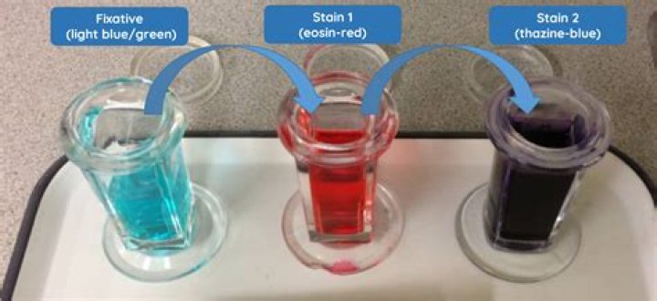
The Diff-Quik stain consists of a fixative agent (methanol, blue), solution I (eosinophilic, orange) and solution II (basophilic, blue). Generally, slides are dipped sequentially into each solution 6 times (or left for 10-15 seconds in each solution), followed by a water rinse and drying.
How do you diff a Quik blood smear?
- Allow smears to dry.
- Dip slide or tape-strip five times, for one second each, into Fixative. …
- Dip slide or tape-strip five times, for one second each, into Stain 1. …
- Dip slide or tape-strip five times, for one second each, into Stain 2. …
- Rinse slide or tape-strip in distilled water or Weise’s buffer, pH 7.2.
Is Diff-Quik a Wright stain?
Diff-Quik® is a commercial stain commonly used in histological staining to rapidly stain and differentiate a variety of smears, commonly blood and vaginal smears. It is based on a modification of the Wright Giemsa stain pioneered by Bernard Witlin in 1970.
How often should you change Diff-Quik stain?
Good laboratory practice should document changing each Diff-Quik stain setup at regular intervals (for example, every week if there is an average of about five evaluations per week). For immediate evaluation on wet fixed samples, an immersion stain setup could pose some threat of cross-contamination.What stains Thiazine?
Chemistry and principle: It is an acidophilic metachromatic dye, which belongs to thiazine group. Therefore, it has a high affinity for acidic compounds; stains the cells / tissues blue with greater nucleic acid content.
What chemical is used first in the diff quick method?
All use methyl alcohol as fixative. What is the fixative made of (the first dipping solution)? 100 WBCs are identified and tallied in the monolayer (the “feathered edge” created by the spreader slide as it draws the blood forward. )
How do you Destain a diff quickly?
Drain excess fluid onto the paper towel. Rinse the slide with the rinse buffer solution or under gently running tap water until the slide is pink and the water runs clear. Rest the slide vertically and allow it to air-dry. Do not wipe or blot the top of the slide with tissue or you will remove or destroy the cells.
Which stain is used for Pap smear?
Papanicolaou stain (also Papanicolaou’s stain and Pap stain) is a multichromatic (multicolored) cytological staining technique developed by George Papanicolaou in 1942. The Papanicolaou stain is one of the most widely used stains in cytology, where it is used to aid pathologists in making a diagnosis.What is Leishman stain procedure?
Leishman Stain is a neutral stain for blood smears which was devised by the British surgeon W. B. Leishman (1865–1926). It consists of a mixture of eosin (an acidic stain), and Methylene blue (a basic stain) in Methyl alcohol and is usually diluted and buffered during the staining procedure.
What magnification do you need to see ear mites?Scan entire slide with 4X or 10X lens for mites or eggs. 1.
Article first time published onHow do you dispose of quick dip stains?
Collect liquid in an appropriate container or absorb with an inert material (e.g. vermiculite, dry sand, earth), and place in a suitable container for reclamation or disposal. Do not use combustible materials, such as sawdust. Do not flush to sewer.
How do you prepare for an ear mite slide?
Put a drop of mineral oil on slide, place tape on slide sticky surface down, and examine under 10X. Ear swabs for mites: Identifies Otodectes cynotis and Demodex mites. Collect debris from ear canal on cotton swab and roll onto slide with a small amount of mineral oil, apply coverslip and examine under 10X.
Is Diff-Quik a Gram stain?
Diff-Quik stain, a variant of Romanowsky stain, is used to quickly identify cells and bacteria. However, it does not differentiate between gram-positive or gram-negative bacteria. … Diff-Quik consists of a fixative (methanol) and eosinophilic (orange) and basophilic (purple) counterstains.
How does Wright stain work?
Wright’s stain is a polychromatic stain consisting of a mixture of eosin and methylene Blue. When applied to blood cells, the dyes produce multiple colors based on the ionic charge of the stain and the various components of the cell.
What is the difference between Wright stain and Giemsa stain?
The main difference between Giemsa stain and Wright stain is that Giemsa stain is used to stain chromosomes to identify chromosome aberrations. But, Wright stain is used to differentiate blood cell types.
What is Thiazine red?
Thiazine red staining is a reliable and rapid pos-mortem diagnostic tool for Alzheimer´s disease.
What is Diff-Quik fixative solution?
RAL Diff-Quik fixative solution. It fixes air-dried blood smears. This methanol based solution will stabilise cellular components. Solution I and II are applied individually to the fixed smear to differentially stain specific cellular components.
What is Basophilic staining?
Basophilic describes the appearance of structures seen in histological sections which take up basic dyes. The structures usually stained are those that contain negative charges, such as the phosphate backbone of DNA in the cell nucleus and ribosomes.
How do you Destain a cytology slide?
Wash slides in deionized or distilled water for 5 min at room temperature. Destain slides in 0.5% HCl in 70% ethanol for 5-15 min at room temperature. Wash slides in deionized or distilled water for 5 min at room temperature.
How do vets stain slides?
- Make sure the sample on the slide is dry.
- Dip the slide in each jar between six-10 times (10-15 seconds in each solution). …
- Wash the slide with water after jar number 3 ONLY.
- Dip the slide in the water jar.
- Never wash the slide in between the staining process.
- Alternative to Jar 4:
What is the difference between Giemsa and Leishman stain?
The key difference between Giemsa Stain and Leishman Stain is that Giemsa staining is useful in the staining of DNA regions of different chromosomes to investigate different aberrations such as translocations and rearrangements, while Leishman stain is useful during blood smear staining and analysis to differentiate …
What are the various stain used commonly in cytology?
The universal stain for cytological preparations is the Papanicolaou stain. Harris’ hematoxylin is the optimum nuclear stain and the combination of OG6 and EA50 give the subtle range of green, blue and pink hues to the cell cytoplasm.
What is the fixative present in Leishman stain?
The fixative present in Leishman stain is Buffered formalin J10% formalin ~Ethy alcohol ~Acetone free methanol 02.
Why is Pap stain used?
Pap stain is a universal stain used for gynecologic and non-gynecologic cytology smear. It is mainly used for oral and cervical cancer screening in asymptomatic population and in the follow up of patients with cancer. Pap test has decreased incidence of cervical cancer by 70% in developed countries.
What is the most critical step in the PAP staining?
Rapid fixation in alcohol (wet version) is essential for pap staining, which brings out nuclear details clearly, allowing better identification of malignant cells.
What does EA 50 stain?
EA50 is a counter stain designed to stain the cytoplasm of cells derived from the human body. The stained cell sample is used to facilitate the diagnosis of the pathological state, and is intended for in-vitro examination of cellular deformity. Cells with high content of keratin are stained yellow.
Can you see Earmites without a microscope?
Ear mites are highly contagious, and animals become infested by direct contact with another infested animal. The mite is barely visible to the naked eye and may be seen as a white speck moving against a dark background.
What do Earmites look like under a microscope?
Ear mites are parasites that live in the ears. Though they are nearly invisible to the naked eye, they look a lot like ticks. Without a microscope, ear mites look like little white dots.
What do rods look like in ear cytology?
Cytology of ear discharge reveals cocci suggestive of Staphylococcus spp. Bacilli appear as elongated rods when viewed under the microscope, they usually appear as single cells. All bacteria and yeast stain dark blue with a Diff Quik stain.
How is ear cytology stained?
Ears: Insert a cotton-tipped applicator into the vertical external ear canal. Then remove the applicator and roll it onto the slide. Heat fix the slide by applying flame to the back of the slide for a few seconds. Stain using the three-part Diff-Quik stain.
How do you swab for ear mites?
If mites are suspected, a second swab with mineral oil applied is used to collect exudate. This material is mixed into a drop of mineral oil on a separate slide. Mite evaluation slides are not stained. Instead, the slide is cover-slipped and examined under the 10× objective for the presence of mites or mite ova.