What are the different parts of the eye called
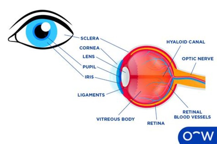
Cornea: This is the front layer of your eye. … Pupil: The pupil is the black dot in the center of your eye that acts as a gateway for light. … Iris: This part is typically referred to as your eye color. … Lens: The lens is behind the iris and pupil.
What are the 15 parts of the eye?
- Parts of the Eye. Here I will briefly describe various parts of the eye:
- Sclera. The sclera is the white of the eye. …
- The Cornea. The cornea is the clear bulging surface in front of the eye. …
- Anterior & Posterior Chambers. The anterior chamber is between the cornea and the iris. …
- Iris/Pupil. …
- Lens. …
- Vitreous Humor. …
- Retina.
What are the 7 structures of the eye?
The main parts of the human eye are the cornea, iris, pupil, aqueous humor, lens, vitreous humor, retina, and optic nerve. Light enters the eye by passing through the transparent cornea and aqueous humor.
What are the 12 parts of the eye?
- The Conjunctiva. The surface of the eye and of the inner eyelids is covered by a clear, protective membrane called the “conjunctiva.” …
- The Sclera. …
- The Cornea. …
- Anterior Chamber. …
- Posterior Chamber. …
- Iris. …
- Pupil. …
- Lens.
What are different parts of the eye and their functions?
The sclera, or white part of the eye, protects the eyeball. The pupil, or black dot at the centre of the eye, is an opening through which light can enter the eye. The iris, or coloured part of the eye, surrounds the pupil. It controls how much light enters the eye by changing the size of the pupil.
What is the white part of your eye called?
The outer layer of the eyeball is a tough, white, opaque membrane called the sclera (the white of the eye). The slight bulge in the sclera at the front of the eye is a clear, thin, dome-shaped tissue called the cornea. The middle layer is the choroid.
Why do we see color?
The human eye and brain together translate light into color. Light receptors within the eye transmit messages to the brain, which produces the familiar sensations of color. … Rather, the surface of an object reflects some colors and absorbs all the others. We perceive only the reflected colors.
What is the corner of your eye called?
Canthus (pl. canthi, palpebral commissures) is either corner of the eye where the upper and lower eyelids meet. More specifically, the inner and outer canthi are, respectively, the medial and lateral ends/angles of the palpebral fissure.How many parts of the eye are there?
The eye itself is made of 10 general components that all work together to keep us seeing well every day.
What is the dark part of the eye called?Your pupil is the black circle in the center of your iris. It regulates how much light enters your eye. Interestingly, the pupil appears black because this tissue absorbs most of the light that passes through it. Your retina is a sensory membrane that covers the entire back surface of your eye.
Article first time published onWhat are rods and cones?
Rods and cones are the receptors in the retina responsible for your sense of sight. They are the part of the eye responsible for converting the light that enters your eye into electrical signals that can be decoded by the vision-processing center of the brain. Cones are responsible for color vision.
What are the two segments of the eye and what is held in each segment?
The anterior segment is divided into two chambers. The front (anterior) chamber extends from the cornea to the iris. The back (posterior) chamber extends from the iris to the lens.
What is the most important part of the eye?
One of the most important parts of the eye is the retina. But why is it so important? Your retina only has one job, but it is a very important one: convert the light that the eye has captured into electric signals that the brain can process.
What colors can dogs see?
Dogs possess only two types of cones and can only discern blue and yellow – this limited color perception is called dichromatic vision.
Can humans see yellow?
Because the human eye has sensors that detect only three color bands as pointed out by S. McGrew and MaxW, it is indeed the case that your brain, retina, and optic nerve are wired to tell you that you are seeing “yellow” when there are no photons at all of that energy entering your eye.
How do we see black?
Objects are visually perceived when they reflect light. A black object does not reflect any light. In other words, no photons are reflected to be detected by the photoreceptors in the retina. A black shape on a colored background appears black because its brightness approaches zero relative to its surroundings.
What is the difference between the sclera and conjunctiva?
The conjunctiva contributes to the tear film and protects the eye from foreign objects and infection. The sclera is the thick white sphere of dense connective tissue that encloses the eye and maintains its shape.
What is a Chemosis?
Chemosis is swelling of the tissue that lines the eyelids and surface of the eye (conjunctiva). Chemosis is swelling of the eye surface membranes because of accumulation of fluid.
Why is my sclera blue?
Blue sclera is caused by a congenitally thinner-than-normal sclera or a thinning of the sclera from disease, which allows the color of the underlying choroidal tissue to show through it.
What is the surface of the eye called?
Covering most of the outside of the eye is a tough white layer called the sclera. A clear thin layer called the conjunctiva covers the sclera. At the very front of the eye is a clear surface, like a window, called the cornea that protects the pupil and the iris behind that window.
What is the part above your eyelid called?
At the top of the upper eyelid is a fold in the skin called a skin crease or the superior palpebral sulcus. It lies around 8 to 11 mm above the margin of the upper eyelid and consists of fibres of the levator aponeurosis. … The inner aspect of the eyelid is called the inner canthal region.
What are the ligaments in the eye?
The suspensory ligament of eyeball (or Lockwood’s ligament) forms a hammock stretching below the eyeball between the medial and lateral check ligaments and enclosing the inferior rectus and inferior oblique muscles of the eye.
What's the pink part of your eye called?
The lacrimal caruncle, or caruncula lacrimalis, is the small, pink, globular nodule at the inner corner (the medial canthus) of the eye.
Why is your pupil black?
The pupil is an opening that lets light into your eye. Since most of the light entering your eye does not escape, your pupil appears black. In dim light, your pupil expands to allow more light to enter your eye.
What is the eyeball Emoji?
👀 Eyes emoji It mostly serves to draw attention to something the user wants to highlight, especially in situations that involve drama and interpersonal tension. It can also be an emoji representation of shifty eyes or the action of side-eyeing. This emoji sometimes appears when someone finds a person attractive.
What is pupil?
Listen to pronunciation. (PYOO-pul) The round opening in the center of the iris (the colored tissue that makes the “eye color” at the front of the eye). The pupil changes size to let light into the eye.
What is the meaning of Emmetropia?
Emmetropia is the refractive state of an eye in which parallel rays of light entering the eye are focused on the retina, creating an image that is perceived as crisp and in focus. Myopia, hyperopia, and astigmatism are abnormalities of this desired condition (Fig. 1-4).
What do ganglion cells do?
Retinal ganglion cells process visual information that begins as light entering the eye and transmit it to the brain via their axons, which are long fibers that make up the optic nerve. There are over a million retinal ganglion cells in the human retina, and they allow you to see as they send the image to your brain.
What is the difference between the anterior and posterior chamber of the eye?
Anterior chamber: This is between your iris and cornea; or the “front” part of the eye. Posterior chamber: This is everything behind the lens of the eye filled with a gel-like transparent fluid.
What is the middle tunic of the eye called?
The middle layer of tissue surrounding the eye, also known as the vascular tunic or „uvea“, is formed – from behind forward – by the choroid, the ciliary body, and the iris. The choroid takes up the posterior five-sixths of the bulb and is mainly comprised of blood vessels.
What divides the eye into anterior and posterior?
The iris divides the eye into the anterior and posterior segments. The pupil can adjust its size independent of the iris.