What are the two parts of the eye
Cornea: This is the front layer of your eye. … Pupil: The pupil is the black dot in the center of your eye that acts as a gateway for light. … Iris: This part is typically referred to as your eye color. … Lens: The lens is behind the iris and pupil.
What is the segment of eye?
The anterior segment refers to the front-most region of the eye, and includes the cornea, iris, and lens. Typically, the phrase “anterior segment surgery” refers to surgery performed on the iris and lens (either natural lens, or synthetic intraocular lens placed during cataract surgery).
How many eye segments are there?
The inside of the eye is divided into three sections called chambers. Anterior chamber: The anterior chamber is the front part of the eye between the cornea and the iris. The iris controls the amount of light that enters the eye by opening and closing the pupil.
What divides the eye into two segments?
The iris divides the eye into the anterior and posterior segments.What are rods and cones?
Rods and cones are the receptors in the retina responsible for your sense of sight. They are the part of the eye responsible for converting the light that enters your eye into electrical signals that can be decoded by the vision-processing center of the brain. Cones are responsible for color vision.
What is the posterior segment of the eye?
The posterior segment of the eye comprises the back two-thirds of the eye, including the vitreous humor, the retina, the choroid and the optic nerve.
What are the major parts of eye and their function?
The sclera, or white part of the eye, protects the eyeball. The pupil, or black dot at the centre of the eye, is an opening through which light can enter the eye. The iris, or coloured part of the eye, surrounds the pupil. It controls how much light enters the eye by changing the size of the pupil.
What separates the anterior and posterior segments of the eye?
Iris: Pigmented tissue lying behind cornea that gives eye color and controls the amount of light entering the eye by varying size of black pupillary opening; separates the anterior chamber from the posterior chamber.What is the anterior and posterior segments of the eye?
The anterior segment is divided into two chambers. The front (anterior) chamber extends from the cornea to the iris. The back (posterior) chamber extends from the iris to the lens. … The back section (posterior segment) extends from the back surface of the lens to the retina.
Does the iris divides the eye into anterior and posterior segments?The iris divides the eye into the anterior and posterior segments. … The lens divides the eye into the anterior segment, located in front of the lens, and the posterior segment, located behind the lens. The pupil can adjust its size independent of the iris.
Article first time published onWhat is the outer corner of the eye called?
Canthus (pl. canthi, palpebral commissures) is either corner of the eye where the upper and lower eyelids meet. More specifically, the inner and outer canthi are, respectively, the medial and lateral ends/angles of the palpebral fissure.
What are the parts of the fibrous layer?
The fibrous layer, which consists of the sclera and cornea. The sclera is an opaque layer which surrounds the posterior five-sixths of the eyeball. The cornea is a transparent layer that is anteriorly continuous with the sclera, occupying the anterior one-sixth of the eyeball.
Where is the limbus of the eye?
The limbus forms the border between the transparent cornea and opaque sclera, contains the pathways of aqueous humour outflow, and is the site of surgical incisions for cataract and glaucoma.
Which layer forms the majority of the cornea?
The majority of the cornea is from the substantia propria. The endothelium is a simple epithelium.
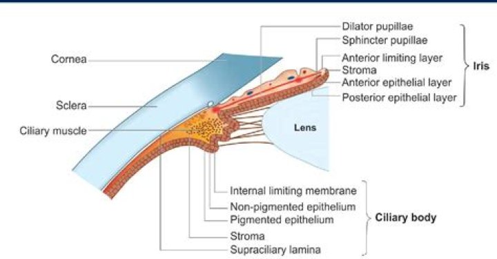
What is the ciliary body of the eye?
A part of the middle layer of the wall of the eye. The ciliary body is found behind the iris and includes the ring-shaped muscle that changes the shape of the lens when the eye focuses. It also makes the clear fluid that fills the space between the cornea and the iris.
What is pupil?
Listen to pronunciation. (PYOO-pul) The round opening in the center of the iris (the colored tissue that makes the “eye color” at the front of the eye). The pupil changes size to let light into the eye.
What is the meaning of Emmetropia?
Emmetropia is the refractive state of an eye in which parallel rays of light entering the eye are focused on the retina, creating an image that is perceived as crisp and in focus. Myopia, hyperopia, and astigmatism are abnormalities of this desired condition (Fig. 1-4).
What is photo receptor?
The photoreceptors are the only cells that can convert incoming light into an electrical signal that can be carried to the brain (via the optic nerve) to create conscious vision.
What is the most important part of the eye?
One of the most important parts of the eye is the retina. But why is it so important? Your retina only has one job, but it is a very important one: convert the light that the eye has captured into electric signals that the brain can process.
What is structure of human eye?
The main parts of the human eye are the cornea, iris, pupil, aqueous humor, lens, vitreous humor, retina, and optic nerve. Light enters the eye by passing through the transparent cornea and aqueous humor. The iris controls the size of the pupil, which is the opening that allows light to enter the lens.
What is the difference between the anterior and posterior chamber of the eye?
The anterior segment or anterior cavity is the front third of the eye that includes the structures in front of the vitreous humour: the cornea, iris, ciliary body, and lens. Within the anterior segment are two fluid-filled spaces: … the posterior chamber between the iris and the front face of the vitreous.
Where is ciliary muscle?
The ciliary muscle is elongated, triangular in shape, and located beneath the anterior sclera just posterior to the limbus. The shortest side of the triangular region faces anterior-inward and it is to this region of the ciliary body that the base of the iris inserts.
What is the vascular layer of the eye?
Middle coat (vascular tunic) The middle layer of tissue surrounding the eye, also known as the vascular tunic or „uvea“, is formed – from behind forward – by the choroid, the ciliary body, and the iris. The choroid takes up the posterior five-sixths of the bulb and is mainly comprised of blood vessels.
What layer contains both a single celled pigmented layer and a neural layer?
Identify the layer that contains both a single-celled pigmented layer and a neural layer. The inner layer (retina) contains both the pigmented layer and the neural layer.
Which of the following is a characteristic of the lens?
Which of the following is a characteristic of the lens? The lens focuses light on the retina.
Which are the characteristics of the cornea?
cornea, dome-shaped transparent membrane about 12 mm (0.5 inch) in diameter that covers the front part of the eye. Except at its margins, the cornea contains no blood vessels, but it does contain many nerves and is very sensitive to pain or touch.
What is temporal Canthus?
Definitions of temporal canthus. the outer corner of the eye. type of: canthus. either of the corners of the eye where the upper and lower eyelids meet.
Why is the outer corner of my eye dry?
When there aren’t enough tears to keep your eyes moist, you can experience dry and itchy eyes, especially in the corners. Dry eyes become more common as you get older because your glands produce fewer tears. Other dry eye triggers include: improper contact lens use.
What is located above the outer corner of the eye?
There is a lacrimal gland just above, and to the outer side, of each eye. The lacrimal glands constantly make a small amount of watery fluid which drains on to the upper part of the eyes. When you blink, the eyelid spreads the tears over the front of the eye.
What is the inner layer of the retina?
Name of the layersContentLayer of PhotoreceptorInner and Outer segments of Photoreceptor
What is a limbus eye?
The limbus forms the border between the transparent cornea and opaque sclera, contains the pathways of aqueous humour outflow, and is the site of surgical incisions for cataract and glaucoma. … The vasculature of the limbus derives in primates primarily from the anterior ciliary arteries.