What are the zones of the bone
These zones include the reserve zone, proliferative zone, and the hypertrophic zone. The reserve zone is the area closest to the secondary center of ossification. It has epiphyseal vessels the pass through this area but do not provide it with oxygen and keeps the oxygen tension low in this area.
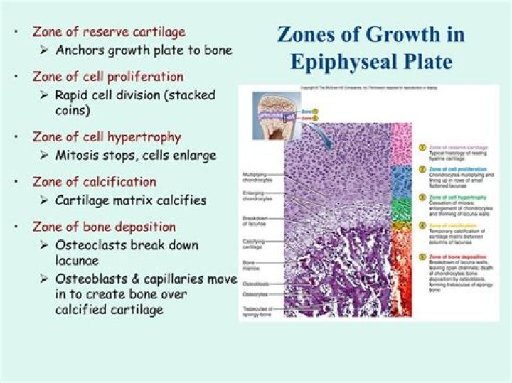
What are the four zones of the epiphyseal plate?
- Proliferation Zone. Zone 1. Cartilage cells undergo mitosis.
- Hypertrophic Zone. Zone 2. Older cartilage cells enlarge.
- Calcification Zone. Zone 3. Matrix becomes calcified; cartilage cells die; matrix begins deteriorating.
- Ossification Zone. Zone 4. New bone formation is occurring.
What are the 6 steps of endochondral ossification?
- Cartilage enlarges; Chondrocytes die.
- blood vessels grow into perichondrium; cells convert to osteoblasts; shaft becomes covered with superficial bone.
- more blood supply and osteoblasts; produces spongy bone; formation spreads on shaft.
- Osteoclasts create medullary cavity; appositional growth.
Where is the zone of ossification?
The epiphyseal plate is the area of growth in a long bone. It is a layer of hyaline cartilage where ossification occurs in immature bones. On the epiphyseal side of the epiphyseal plate, cartilage is formed. On the diaphyseal side, cartilage is ossified, allowing the diaphysis to grow in length.What is the lamellar bone?
Lamellar bone represents the main type of bone in a mature skeleton. It is characterized by an orderly arrangement of collagen bundles and their cells (fig. … The deposited collagen exhibits an orderly lamellar pattern with circular layers of collagen alternating with longitudinal ones.
What is the order of endochondral ossification?
Endochondral ossification begins with mesenchymal tissue transforming into a cartilage intermediate, which is later replaced by bone and forms the remainder of the axial skeleton and the long bones.
What zone of the endochondral ossification is closest to the epiphyseal plate?
The reserve zone is the region closest to the epiphyseal end of the plate and contains small chondrocytes within the matrix. These chondrocytes do not participate in bone growth but secure the epiphyseal plate to the osseous tissue of the epiphysis.
What are the four zones of cartilaginous cells at the epiphyseal plate during endochondral ossification?
- The bone is formed onto a temporary cartilage model.
- The cartilage model grows (zone of proliferation), then chondrocytes mature (zone of maturation) and hypertropy (zone of hypertrophy), and growing cartilage model starts to calcify.
What is Intramembranous and endochondral ossification?
In intramembranous ossification, bone develops directly from sheets of mesenchymal connective tissue. In endochondral ossification, bone develops by replacing hyaline cartilage. Activity in the epiphyseal plate enables bones to grow in length (this is interstitial growth).
Which is true of endochondral ossification?Endochondral ossification occurs within fibrous connective tissue membranes. Endochondral ossification leads to the formation of the clavicles and cranial bones. During endochondral ossification, hyaline cartilage is broken down and replaced with bone. Most bones in the body are formed by intramembranous ossification.
Article first time published onWhat are the zones of the epiphyseal plate quizlet?
- Resting Zone. Area of cartilage on epiphyseal side of epiphyseal plate that is relatively inactive.
- Proliferation (growth) zone. Area of cartilage on diaphysis side of epiphyseal plate that is rapidly dividing. …
- Hypertrophic zone. …
- Calcification zone. …
- Ossification zone.
What cartilages are left in endochondral ossification?
Endochondral ossification involves the replacement of hyaline cartilage with bony tissue. Most of the bones of the skeleton are formed in this manner. These bones are called endochondral bones. In this process, the future bones are first formed as hyaline cartilage models.
Where are the primary and secondary ossification centers located in the long bone during endochondral ossification?
Each of these bones has a primary center of ossification. The zone of endochondral ossification spreads from the primary ossification center toward the ends of the cartilage. These slides do not show secondary ossification centers. Note the bone of the diaphysis.
Where is the primary center of ossification in endochondral ossification of a long bone quizlet?
In long bones the primary centers occur in the diaphysis/shaft and in irregular bones the primary centers occur usually in the body of the bone. The site where bone formation continues after beginning in the long shaft or body of the bone, usually in an epiphysis; secondary ossification center.
How many steps are in endochondral ossification?
Figure 6.13. Endochondral Ossification Endochondral ossification follows five steps. (a) Mesenchymal cells differentiate into chondrocytes. (b) The cartilage model of the future bony skeleton and the perichondrium form.
What are the stages of ossification?
The process of bone formation is called osteogenesis or ossification. After progenitor cells form osteoblastic lines, they proceed with three stages of development of cell differentiation, called proliferation, maturation of matrix, and mineralization.
What is the canaliculi in bone?
Bone canaliculi are microscopic canals between the lacunae of ossified bone. The radiating processes of the osteocytes (called filopodia) project into these canals. … Materials picked up by osteocytes adjacent to blood vessels are distributed throughout the bone matrix via the canaliculi.
Is trabecular bone lamellar?
A single trabecula is composed of lamellar tissue with osteocytes lying in lacunae with a network of canaliculi similar to that of the cortical tissue.
What is haversian Canal?
Haversian canals are a series of tubes around narrow channels formed by lamellae. The Haversian canals surround blood vessels and nerve fibers throughout the bone and communicate with osteocytes. The canals and the surrounding lamellae are called a Haversian system (or an osteon).
What are the four main steps of the bone ossification process?
There are four stages in the repair of a broken bone: 1) the formation of hematoma at the break, 2) the formation of a fibrocartilaginous callus, 3) the formation of a bony callus, and 4) remodeling and addition of compact bone.
What are primary and secondary centers of ossification?
The primary ossification center is the first place where the bone formation begins in the axle of a long bone or in the body of an irregular bone. Conversely, the secondary ossification center is the area of ossification that appears after the primary center of ossification at the epiphysis of edges of bones.
What are the 5 steps of ossification in a long bone?
- hyaline cartilage is completly covered with bone matrix by osteoblasts. …
- cartilage is digested away by osteoclasts, opening up a medullary cavity within the bone. …
- blood vessels, neves, and osteoblasts, and osteoclasts, invade the internal shaft and spongy bone is formed.
What are the main differences between dermal and endochondral ossification?
Ossification is different from calcification. Ossification takes place approximately six weeks after fertilization in an embryo. Dermal ossification is the development of bone from fibrous membranes, while endochondral ossification is a type of development of bone from hyaline cartilage.
Which statement about endochondral ossification is false?
False; Endochondral ossification requires the formation of a hyaline cartilage model; flat bones do not form this way. Osteopenia is an adult-onset reduction in bone mass that can lead to osteoporosis.
Which of the following describes the zone of calcification in an epiphyseal plate?
Which of the following describes the zone of calcification in an epiphyseal plate? Minerals are deposited between the columns of lacunae.
What happens in the zone of resting cartilage?
Our findings suggest that resting zone cartilage makes important contributions to endochondral bone formation at the growth plate: 1) the resting zone contains stem-like cells that give rise to clones of proliferative chondrocytes; 2) the resting zone produces a GPOF, a morphogen that directs the alignment of the …
What occurs in the zone of resting cartilage?
Zone of Resting Cartilage: This is the area where the hyaline cartilage remains unchanged. … Zone of Calcified Cartilage: In this area, cartilage is calcified into bone tissue and blood vessels form. This happens at the junction between the epiphysis and diaphysis.
What is the primary ossification center in endochondral ossification?
Primary center of ossification The perichondrium becomes the periosteum. The periosteum contains a layer of undifferentiated cells (osteoprogenitor cells) which later become osteoblasts. The osteoblasts secrete osteoid against the shaft of the cartilage model (Appositional Growth).