What does the vestibular nerve do
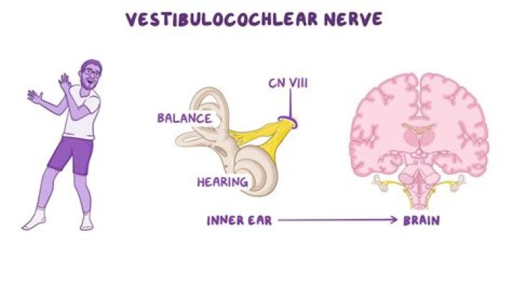
The vestibulocochlear nerve sends balance and head position information from the inner ear (see left box) to the brain.
How do the auditory and vestibular systems differ from one another?
The vestibular system, which is not auditory, detects linear acceleration and angular acceleration and deceleration. Both the auditory system and vestibular system use hair cells as their receptors. Auditory stimuli are sound waves.
Where do the cochlear and vestibular nerves pass through?
Cranial nerves The vestibulocochlear nerves originate in the monitoring receptors of the internal ear—the vestibule and cochlea. They pass through the internal acoustic meatus of the temporal bones, ending in the vestibular and cochlear nuclei of the pons and medulla oblongata.
What nerve does the vestibular and cochlear nerve create?
Vestibulocochlear nerveInferior view of the human brain, with the cranial nerves labelled.DetailsToCochlear nerve, vestibular nerveInnervatesHearing, balanceWhat nerve innervates the cochlea?
The vestibulocochlear nerve, also known as cranial nerve eight (CN VIII), consists of the vestibular and cochlear nerves.
Is the cochlea part of the vestibular system?
The main peripheral component of the vestibular system is an elaborate set of interconnected canals—the labyrinth—that has much in common, and is in fact continuous with, the cochlea.
Is cochlea part of nervous system?
The cochlear nerve (also auditory nerve or acoustic nerve) is one of two parts of the vestibulocochlear nerve, a cranial nerve present in amniotes, the other part being the vestibular nerve. The cochlear nerve carries auditory sensory information from the cochlea of the inner ear directly to the brain.
In what ways are the vestibular and auditory sense organs similar?
In what ways are the vestibular and auditory sense organs similar? Answer: Both the vestibular and auditory system have elaborate set of interconnected chambers that use endolymph movement and specialized hair cells to detect stimuli. 2.What does the cochlea contain?
Structure of the cochlea. The cochlea contains the sensory organ of hearing. It bears a striking resemblance to the shell of a snail and in fact takes its name from the Greek word for this object. The cochlea is a spiral tube that is coiled two and one-half turns around a hollow central pillar, the modiolus.
Where does the vestibular nerve start?The vestibular portion of the vestibulocochlear nerve originates in a group of nerve cells called the vestibular ganglion, in the internal acoustic meatus, a channel in the temporal bone through which the facial and auditory nerves and some blood vessels run.
Article first time published onWhat nucleus of vestibulocochlear nerve is cochlear?
Nuclei. There are two special sensory cochlear nuclei and four special sensory vestibular nuclei located within the lower pons and upper medulla.
Which of the cranial nerves transmits from the cochlea and semicircular canals to the brain?
These semi-circular canals are filled with fluid and have some small calcium crystals embedded in the lining. Coming from the inner ear and running to the brain is the eighth cranial nerve, the auditory nerve. This nerve carries both balance and hearing information to the brain.
How many vestibular nerves are there?
The vestibular nerve enters the brain stem at the pontomedullary junction and contains two divisions, the superior and inferior vestibular nerves. The superior vestibular nerve innervates the utricle, as well as the superior and lateral canals.
Is the vestibulocochlear nerve the auditory nerve?
vestibulocochlear nerve, also called Auditory Nerve, Acoustic Nerve, or Eighth Cranial Nerve, nerve in the human ear, serving the organs of equilibrium and of hearing.
Where does the vestibular nerve project to in the brain?
The vestibular labyrinth is made up of the semicircular canals and the otolith organs (all discussed below), and contains receptors for vestibular sensations. These receptors send vestibular information via the vestibulocochlear nerve to the cerebellum and to nuclei in the brainstem called the vestibular nuclei.
Is the Vestibulocochlear nerve sensory or motor?
No.NameSensory, motor, or bothVTrigeminalBoth sensory and motorVIAbducensMainly motorVIIFacialBoth sensory and motorVIIIVestibulocochlear In older texts: auditory, acoustic.Mostly sensory
What part of the ear contains receptors for the vestibular branch of the auditory nerve?
There are five vestibular receptor organs in the inner ear: the utricle, the saccule, and three semicircular canals.
What is a basic vestibular evaluation?
Vestibular testing involves a series of tests that are administered when you are experiencing dizziness. They are used to determine whether symptoms of dizziness are being caused by the balance system of the inner ear.
What is a cochlear nerve?
The cochlear nerve is primarily responsible for transmitting the electrical impulses generated for hearing and localization of sound. The nerve has its origin in the bipolar cells of the spiral ganglion of the cochlea, which is located adjacent to the inner margin of the bony spiral lamina.
Where is the cochlea in the ear?
While the cochlea is technically a bone it plays a vital role in the function of hearing rather than simply being another component of the skeletal system. It is located within the inner ear and is often described as hollow and snail- or spiral-shaped.
What is the cochlea known as?
The cochlea is the part of the inner ear involved in hearing. It is a spiral-shaped cavity in the bony labyrinth, in humans making 2.75 turns around its axis, the modiolus. … The name cochlea derives from Ancient Greek κοχλίας (kokhlias) ‘spiral, snail shell’.
How can you tell the difference between peripheral and central vestibular disorders?
As a result, the head tilts towards the loss of muscle tone. In cases with peripheral vestibular lesion, head tilt is towards the side of the lesion. For example, left inner ear disease (i.e., otitis media interna) causes left head tilt. A unilateral central vestibular lesion can cause a head tilt to either side.
What is the vestibular sense how does it involve the semicircular canals and vestibular sacs?
Gravitation and movement sensations are produced by movement of two vestibular sacs in each ear that lie between the semicircular canal and the cochlea. Both sacs are filled with millions of tiny crystals that bend hair cells when moved. In turn, impulses giving a sense of position are sent to the brain.
What is the difference between cochlea and cochlear duct?
The cochlear duct is part of the cochlea. It is separated from the tympanic duct (scala tympani) by the basilar membrane. It is separated from the vestibular duct (scala vestibuli) by the vestibular membrane (Reissner’s membrane). The stria vascularis is located in the wall of the cochlear duct.
What does the cochlea resemble?
The cochlea is a coiled structure that resembles a snail shell (cochlea comes from the Greek kochlos, which means “snail”); it is found within the inner ear.
What is the main function of the cochlea?
The cochlea: function and anatomy The cochlea has a very important function in the hearing process: In the cochlea, It transforms sound waves into electrical impulses which are sent on to the brain. The brain then translates the impulses into sounds that we know and understand.
How the vestibular apparatus enables the brain to interpret the body's position and movements?
Structure of the vestibular receptors. The vestibular receptors lie in the inner ear next to the auditory cochlea. They detect rotational motion (head turns), linear motion (translations), and tilts of the head relative to gravity and transduce these motions into neural signals that can be sent to the brain.
What is part of the vestibular system?
vestibular system, apparatus of the inner ear involved in balance. The vestibular system consists of two structures of the bony labyrinth of the inner ear, the vestibule and the semicircular canals, and the structures of the membranous labyrinth contained within them.
What is the macula in the vestibular system?
Vestibular structures Each sac has on its inner surface a single patch of sensory cells called a macula, which monitors the position of the head relative to the vertical.
Is vestibular nerve ipsilateral?
In summary, remember that the lateral vestibulo-spinal tract is ipsilateral and long; the medial vestibulo-spinal tract is bilateral but shorter. The main ascending tracts are from the superior and medial vestibular nuclei to the extraocular muscles through the medial longitudinal fasciculus (MLF).
What part of the vestibular apparatus connects directly to the cochlear duct?
Saccule. The saccule is an almost globular-shaped sac that lies in the spherical recess on the medial wall of the vestibule. It is connected anteriorly to the cochlear duct by the ductus reuniens and posteriorly to the endolymphatic duct via the utriculosaccular duct.