What is a CT petrous bones scan for
The CT is a particularly powerful exam for studying the five bony parts of the T-bone: Squamous: forms the lateral wall of the middle cranial fossa. Mastoid. Petrous: contains the inner ear, internal auditory canal (IAC), petrous apex. Tympanic: forms most of the bony external ear.
What is the temporal bone CT scan and what does it show?
Temporal bone CT is a limited kind of head CT that focuses on the lower part of the skull and the surrounding soft tissues, and is often used in patients with hearing loss, chronic ear infections, and middle and inner ear diseases.
What conditions can a CT scan detect?
CT scans can detect bone and joint problems, like complex bone fractures and tumors. If you have a condition like cancer, heart disease, emphysema, or liver masses, CT scans can spot it or help doctors see any changes. They show internal injuries and bleeding, such as those caused by a car accident.
What does the petrous bone mean?
Definition of petrous : of, relating to, or constituting the exceptionally hard and dense portion of the human temporal bone that contains the internal auditory organs.Does a CT scan show a tumor?
CT scans can show a tumor’s shape, size, and location. They can even show the blood vessels that feed the tumor – all without having to cut into the patient. Doctors often use CT scans to help them guide a needle to remove a small piece of tissue. This is called a CT-guided biopsy.
How long does a temporal bone CT take?
Depending on the reason for your test, the procedure can take anywhere from 10 to 30 minutes. You will receive the results of the exam from your doctor.
What is found in the temporal bone?
The temporal bone contributes to the lower lateral walls of the skull. It contains the middle and inner portions of the ear, and is crossed by the majority of the cranial nerves. The lower portion of the bone articulates with the mandible, forming the temporomandibular joint of the jaw.
What are processes of petrous part?
The petrosal process is a sharp process below the notch for the passage of the abducent nerve on either side of the dorsum sellae of the sphenoid bone. It articulates with the apex of the petrous portion of the temporal bone, and forms the medial boundary of the foramen lacerum.What is the function of the petrous part?
It transmits the facial and acoustic nerves and the internal auditory branch of the basilar artery. The lateral end of the canal is closed by a vertical plate, which is divided by a horizontal crest, the falciform crest, into two unequal portions.
Which nerve passes close to the petrous part of the temporal bone?The vagus nerve, by its auricular branch that separates from it just beneath the cranial base. It then runs in an osseous canal nestled in the petrous part of the temporal bone (mastoid canaliculus) and gives off a small branch to the facial nerve.
Article first time published onShould I be worried about having a CT scan?
What Are the Risks? CT scans do use radiation that can cause effects in living tissue, however this level of radiation is monitored very closely. Aside from radiation, which we will dive into shortly, the only other risk is a false positive that may lead to unnecessary follow-up tests.
Can you have a CT scan and a bone scan the same day?
Bone Scan, Skeletal Imaging If a CT scan is scheduled for the same day, an IV line will be placed prior to the injection for the bone scan. You may not eat until after the CT scan is completed.
Why would a doctor order a full body scan?
Why doctors order full body CT scans Detect internal injuries and bleeding. Find blood clots, tumors, and infections. Show bone fractures and muscle inflammation. Monitor diseases of the heart, liver, and lungs.
What if my CT scan is abnormal?
CT scan results are considered normal if the radiologist didn’t see any tumors, blood clots, fractures, or other abnormalities in the images. If any abnormalities are detected during the CT scan, you may need further tests or treatments, depending on the type of abnormality found.
Would a CT scan show a blood clot?
CT scans detect and diagnose blood clots by providing detailed, accurate imagery of the body’s blood vessels and their obstructions. Doctors generally use two CT scan techniques for blood clot detection and diagnosis — CT venography and CT pulmonary angiography.
How small a tumor can a CT scan detect?
Due to the physical limitations, however, the minimum lesion size that can be measured with CT is about 3 mm (24). Modern MR imaging systems demonstrate similar lesion detection limits (25).
Why is the temporal bone important?
The temporal bone is a thick, hard bone that forms part of the side and base of the skull. This bone protects nerves and structures in the ear that control hearing and balance.
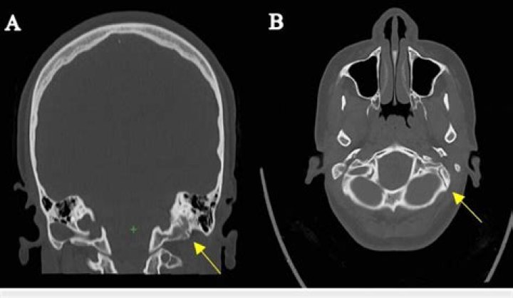
What does a temporal bone fracture feel like?
Temporal bone fractures, especially the oblique variety (see above), may impair hearing and cause dizziness. There often is blood seen behind the ear-drum (hemotympanum). Either a conductive or sensorineural hearing loss or both may be present.
What muscles are attached to the temporal bone?
Muscular attachments The temporalis muscle originates from the temporal fossa, which is formed partially by the lateral aspect of the temporal bone. The sternocleidomastoid, splenius capitis, longissimus capitis and digastric are all attached to the mastoid process of the temporal bone.
Can a CT scan detect inner ear problems?
Computerized Tomography (CT) Scan A CT scan is often used to let doctors see abnormalities, such as fractures or thinning bone, around the inner ear. Vision Tests Sometimes vision tests are recommended to help doctors find a cause for vertigo symptoms.
What's better CT or MRI?
Both MRIs and CT scans can view internal body structures. However, a CT scan is faster and can provide pictures of tissues, organs, and skeletal structure. An MRI is highly adept at capturing images that help doctors determine if there are abnormal tissues within the body. MRIs are more detailed in their images.
Why would a fracture in the anterior temporal bone?
Temporal bone trauma is usually the result of blunt head injury and patients commonly suffer from multiple other body injuries. Motor vehicle accidents are the most common cause, with falls and gunshot wounds contributing to a lesser extent.
Why is it called petrous part of temporal bone?
The petrous and mastoid parts of the temporal bone, which derive from the periotic bone, formed from the fusion of a number of bones surrounding the ear of reptiles.
What type of bone is the sphenoid bone?
The sphenoid is an unpaired bone. It sits anteriorly in the cranium, and contributes to the middle cranial fossa, the lateral wall of the skull, and the floor and sides of both orbits. It has articulations with twelve other bones: Unpaired bones – Occipital, vomer, ethmoid and frontal bones.
What is frontal bone?
Frontal bone: The large bone that makes up the forehead and supplies the upper edge and roof of the orbit (eye socket). The frontal bone articulates (comes together) with a number of other bones including the parietal, nasal, ethmoid, maxillary, and zygomatic bones.
Is the occipital bone a flat bone?
Occipital bone. This is a flat bone located in the very back of your skull. It has an opening that allows your spinal cord to connect to your brain.
What is the most dense bone in the human body?
The human petrous bone in the skull protects the inner ear structures. Though it is one of the hardest, densest bones in the body, some portions (such as the area in orange, protecting the cochlea) are denser than others.
Where is the petrous ridge located?
On the interior of the skull, the petrous portion of each temporal bone forms the prominent, diagonally oriented petrous ridge in the floor of the cranial cavity. Located inside each petrous ridge are small cavities that house the structures of the middle and inner ears.
What is the weakest part of the skull?
Clinical significance The pterion is known as the weakest part of the skull. The anterior division of the middle meningeal artery runs underneath the pterion. Consequently, a traumatic blow to the pterion may rupture the middle meningeal artery causing an epidural haematoma.
What does the mastoid process do?
Mastoid Process Function The mastoid process’ main function is to provide an area of attachment to several important muscles in the head. For instance, it is the attachment site of certain muscles of the neck: Sternocleidomastoid muscle – enables the rotation of the head to the contralateral side.
What is the most common reason for a CT scan?
Chronic back pain or an injury to the spine are among the most common reasons to have a CT scan. A doctor may also order a spinal CT scan to: Evaluate spinal fractures. Assess the condition of the spine before and after surgical procedures.