What is a digital fluoroscopy
Digital fluoroscopy is a form of x-ray used to examine tissues and deep body structures. There are procedures done with digital fluoroscopy which may require the patient to drink a safe liquid called barium. The procedure is often used to look at the internal organs that play a part in swallowing and digestion.
What is fluoroscopy and how does it work?
Fluoroscopy is a study of moving body structures–similar to an X-ray “movie.” A continuous X-ray beam is passed through the body part being examined. The beam is transmitted to a TV-like monitor so that the body part and its motion can be seen in detail.
What are the advantages of digital fluoroscopy?
Advantages to this technology include: Faster turn-around times and immediate image preview. Ability to digitally transfer images to other systems for ease of analysis. Increased image enhancement and detail.
What is the difference between conventional fluoroscopy and digital fluoroscopy?
Conventional and digital fluoroscopy differ primarily in the imaging system, i.e., an image intensifier-video camera system versus a digital imaging chain which may have neither an image intensifier nor video camera. In general, all other portions of the equipment are similar.What are the advantages of digital fluoroscopy over conventional fluoroscopy?
Introduction: Digital fluoroscopy (DF) advantages over conventional fluoroscopy include: Speed of image acquisition. Ability to post process (enhance image contrast). Lower patient dose.
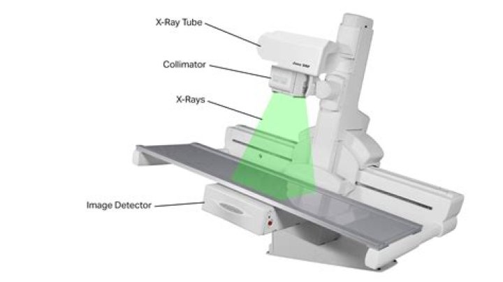
What are the components of fluoroscopy?
The key components include an X-ray tube, spectral shaping filters, a field restriction device (aka collimator), an anti-scatter grid, an image receptor, an image processing computer and a display device.
How is fluoroscopy different from radiography?
Radiography or X-ray and fluoroscopy procedures seem similar. However, fluoroscopy obtains moving images of the inner part of the body and radiography uses gamma rays to develop a static image of the internal structure of a body.
What dye is used in fluoroscopy?
Types of dyes used include: Barium sulfate, a white-chalky substance. Water-soluble agents. Omnipaque (iohexol)What is a fluoro injection?
Fluoroscopically-guided injections are a non-operative, conservative method of treatment for ongoing and chronic back pain or joint pain. These injections are outpatient procedures that can help relieve pain quickly by reducing inflammation in the affected area.
What is a major disadvantage of direct fluoroscopy?Radiation-related risks associated with fluoroscopy include: radiation-induced injuries to the skin and underlying tissues (“burns”), which occur shortly after the exposure, and. radiation-induced cancers, which may occur some time later in life.
Article first time published onWhat are the two methods of flat panel image capture?
There are two types of flat panel detectors used in digital radiography: direct and indirect. One type of detector is based on a direct conversion process of x-rays while other one uses an indirect conversion method. The term direct refers to the acquisition and capture of x-ray images without user intervention.
Why is digital radiography better?
With the addition of computer technology, digital radiography has become a much more efficient, cost effective, and an even safer method of producing diagnostic images. … While traditional X-rays are considered safe, digital X-rays produce 80% less radiation than traditional.
What are the advantages of digital imaging?
The Benefits of Digital Imaging and Impressions Impressions are stored electronically for easy access. Issues may be diagnosed immediately, as no time is needed for processing images. Precise imaging allows for improved fit of restorations. Saves time for the patient and dentist.
What are the principal advantages of digital radiography over conventional radiography?
Advantages over Conventional Radiography Images produced with conventional radiography have limited dynamic range, but digital radiography allows for many exposures to build up a high contrast image. A digital radiograph can be easily copied, e-mailed, and enhanced without degradation.
How much radiation do you get from fluoroscopy?
Getting a fluoroscopic procedure exposes a patient to as much radiation as 250 to 3,500 chest X-rays. For perspective, a person gets the equivalent of one chest X-ray from normal background radiation in about two and a half days.
How much does a fluoroscopy machine cost?
LowHighAverage$250,204$621,347$395,085.90
What is pulsed fluoroscopy?
Pulsed Fluoroscopy Instead of using continuous x-ray tube current, some systems create a short pulse of x-rays at the beginning of each frame, delivering the same dose per frame. For instance, if 3 mA is continuously on for 30 frames/second (frame/s) imaging, the effective mAs is 3 mA/30 frame/s = 0.1 mAs /frame.
How does fluoroscopy help in diagnosis?
Fluoroscopy allows your doctor to see your organs and tissues working on a video screen, similar to watching a movie. Fluoroscopy helps diagnose and treat many conditions of the blood vessels, bones, joints, and digestive, urinary, respiratory and reproductive systems.
Why fluoroscopy is used in radiology?
Coronary angiography is an example of a fluoroscopy procedure. A small tube (catheter) is inserted into an artery of the heart. Contrast dye moves through the catheter into the blood vessels. The fluoroscopy shows how the blood moves through the vessels and allows the healthcare provider to locate any blockages.
How is the fluoroscopic image formed?
In radiography, an image intensifier is simply a device which amplifies the visible light resulting from the fluoroscopic process. … At the photocathode, light energy is used to promote the energy of existing electrons within the material so that they are emitted from it.
What is 3D fluoroscopy?
This new technology allows the use of fluoroscopy coregistered with a 3D data set reconstructed from the acquired attenuation information. The needle trajectory is planned in the 3D data set using the needle path–planning software. The calculated trajectory is then projected on to the real-time fluoroscopy image.
Why is it called fluoroscopy?
Both live moving images and recorded still images were available from the beginning with simple equipment; thus, both “looking with a fluorescent screen” (fluoro- + -scopy) and “recording/engraving with radiation” (radio- + -graphy) were immediately named with New Latin words—both words are attested since 1896.
What is a fluoroscopy Esophagram?
A barium swallow test (cine esophagram, swallowing study, esophagography, modified barium swallow study, video fluoroscopy swallow study) is a special type of imaging test that uses barium and X-rays to create images of your upper gastrointestinal (GI) tract.
How long does a fluoroscopy procedure take?
This exam is usually completed within 20 minutes. If a Small Bowel Exam is also performed, then the exam could take up to 4hrs.
What is fluoro guidance?
Fluoroscopy is a form of X-ray imaging guidance that helps your doctor to locate the internal injection site where an injection, such as a steroid or joint injection is to be administered for pain relief. Fluoroscopy is like GPS (global positioning system) navigation for the tip of an injection needle.
How long does a fluoroscopic injection last?
The steroid lasts for up to six weeks but the effects of the injection can sometimes last much longer. Your doctor will normally arrange a follow-up appointment with you after your injection. There are small risks associated with this injection, which you should be aware of before you proceed.
Does contrast hurt?
It’s kind of like a dye in the way that it temporarily changes how your insides appear on a medical image, but it won’t change the color of anything and it won’t hurt you. You might need contrast when you are having an X-ray, CT, MRI, or ultrasound exam.
Can you eat or drink before a fluoroscopy?
Don’t drink or eat anything for four hours before this procedure. Let the doctor know if you have allergies in general and an allergy to contrast dye or iodine. This injection does require contrast material containing iodine.
How fluoroscopy are produced?
Fluoroscopy uses a mobile x-ray source that produces x-rays continuously or in short pulses. The x-ray beam is focused on the patient in a relatively small field of view and is detected by a device called an image intensifier, which projects the resulting image on a monitor as a real-time image.
What is C arm fluoroscopy?
C-Arm is a mobile imaging unit used primarily for fluoroscopic imaging during surgical and orthopedic procedures. It also consists of a computer workstation used to view, manipulate, store and transfer the images.
Who can operate a fluoroscopy machine?
Some state radiation safety regulations require fluoroscopic equipment operators to obtain special permits to perform fluoroscopy. For example, in the state of California, a radiologic technologist or a physician assistant must hold a California fluoroscopic permit to participate in fluoroscopic examinations [5,6].