What is fluorescence staining
fluor·es·cent stain a stain or staining procedure using a fluorescent dye or substance that will combine selectively with certain tissue components and that will then fluoresce upon irradiation with ultraviolet or violet-blue light.
What does the auramine-rhodamine do?
The auramine-rhodamine staining technique being a histological type of stain that is used to stain and demonstrate the presence of Acid Fast-bacilli under a fluorescent Microscope and also known as Truant auramine–rhodamine stain, demonstrates the anatomy of the bacterial bacilli-cell.
What does Ziehl Neelsen stain?
Ziehl–Neelsen staining is a bacteriological stain used to identify acid-fast organisms, mainly Mycobacteria. It is named for two German doctors who modified the stain: the bacteriologist Franz Ziehl (1859–1926) and the pathologist Friedrich Neelsen (1854–1898).
What is auramine O stain?
Our Auramine O Stain is a fluorochrome stain used in the microscopic detection and examination of acid-fast mycobacteria. Acid-fast organisms have cell walls that are resistant to conventional staining by aniline dyes such as the Gram stain.What is the immunofluorescence technique?
Direct immunofluorescence technique: it is a one-step histological staining procedure for identifying in vivo antibodies that are bound to tissue antigens, using a single antibody labeled with a fluorophore [5] for staining the tissues or cells. The antibody recognizes the target molecule and binds to it.
What is auramine used for?
Auramine colourants are used for dyeing of leather, jute, tanned cotton, and paints, and as dye components in inking ribbons, ballpoint pastes, oils and waxes, and carbon paper. The most important applications are in paper dyeing and flexographic printing (IARC, 2010).
What are stains used for in microbiology?
Gram staining is used to determine gram status to classifying bacteria broadly based on the composition of their cell wall. Gram staining uses crystal violet to stain cell walls, iodine (as a mordant), and a fuchsin or safranin counterstain to (mark all bacteria).
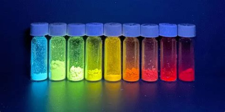
What are Fluorochromes give an example?
Examples of fluorochromes used in the detection of art materials are: Berberine sulfate, Acridine orange, Acridine yellow, Auramine O, Blancophor R, Cycloheptaamylose dansyl chloride, Dichlorofluorescein, Fluorescein isothiocyanate, Lissamine Rhodamine B Sulfonyl Chloride, Primuline, Pyronine Y, Rhodamine B, Rosaniline …What is the principle application of negative staining?
The main purpose of Negative staining is to study the morphological shape, size and arrangement of the bacteria cells that is difficult to stain. eg: Spirilla. It can also be used to stain cells that are too delicate to be heat-fixed. It is also used to prepare biological samples for electron microscopy.
How do you make auramine stain?- Place the fixed smear on a staining rack and flood slide with rhodamine-auramine for 15 minutes. …
- Wash off the stain with distilled water.
- Flood slide with fluorescent decolorizer (i.e acid-alcohol) for 2-3 minutes.
- Rinse thoroughly with distilled water.
- Flood slide with potassium permanganate for 3-4 minutes.
What is the purpose of adding Calcofluor white?
Calcofluor White stain can be used in Clinical Mycology and Parasitology: to demonstrate the presence of amoebic parasites by identifying their cysts. stain bud scars of yeast cells with a higher content of chitin enabling quantification of the bud scars which indicates the age of the cell.
What is meant by acid fast bacilli?
Acid-fast bacillus (AFB) is a type of bacteria that causes tuberculosis and certain other infections. Tuberculosis, commonly known as TB, is a serious bacterial infection that mainly affects the lungs.
Why is the Ziehl-neelsen method called the hot method?
The Ziehl-Neelsen method of staining is also called the hot method as it involves heating the carbolfuchsin stain. In contrast, the historic method of staining called the Kinyoun method does not involve heating and is hence known as the cold method.
Which condition is identified by Ziehl-neelsen method?
Conventional smear microscopy with the Ziehl-Neelsen (ZN) stain is a rapid and practical method for detecting acid-fast bacilli (AFB), especially in low-income countries, due to its rapidity, low cost, and high positive predictive value for tuberculosis (14).
Why is the Ziehl-neelsen considered a hot method?
Mycobacteria, which do not stain well by Gram stain, are stained with carbol fuchsin combined with phenol. In the ‘hot’ ZN technique, the phenol-carbol fuchsin stain is heated to enable the dye to penetrate the waxy mycobacterial cell wall.
What is immunofluorescence and its types?
Immunofluorescence (IF) is a type of immunohistochemistry technique that utilizes fluorophores to visualize various cellular antigens such as proteins.
What is immunofluorescence microbiology?
Immuno Fluorence is defined as various techniques used for detecting an antigen or antibody in a sample by coupling its specifically interactive antibody or antigen to a fluorescent dye/compound, mixing with the sample, and then observing the reaction under an ultraviolet-light fluorescence microscope.
What is the importance of immunofluorescence?
Immunofluorescence (IF) is an important immunochemical technique that allows detection and localization of a wide variety of antigens in different types of tissues of various cell preparations.
What is a staining technique?
Staining is an auxiliary technique used in microscopy to enhance contrast in the microscopic image. Stains and dyes are frequently used in biology and medicine to highlight structures in biological tissues for viewing, often with the aid of different microscopes.
What is the purpose of staining?
The main purpose of staining is to highlight cells and parts of cells. Over 20 different types of stains exist, and the type of stain you use depends on what you are looking for.
What is simple staining technique?
Simple staining involves directly staining the bacterial cell with a positively charged dye in order to see bacterial detail, in contrast to negative staining where the bacteria remain unstained against a dark background.
How do you make auramine rhodamine stain?
- Place the “fixed” smear on a staining rack and flood slide with rhodamine-auramine for 15 minutes. …
- Wash off the stain with distilled water.
- Flood slide with fluorescent decolorizer for 2-3 minutes.
- Rinse thoroughly with distilled water.
- Flood slide with potassium permanganate for 3-4 minutes.
How does acid-fast stain work?
Some of the sample is placed on a glass slide, stained, and heated. The cells in the sample hold onto the dye. The slide is then washed with an acid solution and a different stain is applied. Bacteria that hold onto the first dye are considered “acid-fast” because they resist the acid wash.
What are the stains used in negative staining?
For bright-field microscopy, negative staining is typically performed using a black ink fluid such as nigrosin and India ink. The specimen, such as a wet bacterial culture spread on a glass slide, is mixed with the negative stain and allowed to dry.
What is an example of negative stain?
In a negative staining technique, an acidic, anionic dye is mixed with a cell sample. … India ink is the classic example of a negative stain. It will turn the background a dark brown to black, leaving the clear, bright cells unstained and highly visible.
What is positive and negative staining?
Alternatively, positive and negative staining techniques can be combined to visualize capsules: The positive stain colors the body of the cell, and the negative stain colors the background but not the capsule, leaving halo around each cell.
What is the difference between a fluorochrome and fluorophore?
As nouns the difference between fluorochrome and fluorophore is that fluorochrome is any of various fluorescent dyes used to stain biological material before microscopic examination while fluorophore is (biochemistry) a molecule or functional group which is capable of fluorescence.
What is the meaning of fluorochrome?
Definition of fluorochrome : any of various fluorescent substances used in biological staining to produce fluorescence in a specimen.
What can fluorochrome detect?
The chemical properties of the fluorochrome determine whether its electrons can be excited to the higher energy state by a specific wavelength of laser light.
What is sputum smear?
A sputum stain for Mycobacteria is a laboratory test performed on a sample of your sputum, or phlegm. It’s also known as an acid-fast bacillus (AFB) stain or a tuberculosis (TB) smear. A doctor typically orders the test to determine if a person has tuberculosis (TB) or another type of mycobacterial infection.
What does AFB mean in medical terms?
Acid- Fast Bacilli (AFB) smear and culture are two separate tests always performed together at the MSPHL, Tuberculosis (TB) Unit. AFB smear refers to the microscopic examination of a fluorochrome stain of a clinical specimen.