What is petrous bone
The human petrous bone in the skull protects the inner ear structures. … Found near our ears, this pyramid-shaped portion of the temporal bone is nicknamed the petrous bone.
Where is the petrous bone located?
The petrous part is a wedge shaped mass of bone located between the sphenoid and occipital bones within the cranial cavity. It is the most medial part of the temporal bone, and it is the landmark dividing the middle and posterior cranial fossae from each other.
Is the petrous bone part of the skull?
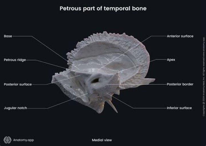
The petrous part of the temporal bone is pyramid-shaped and is wedged in at the base of the skull between the sphenoid and occipital bones. Directed medially, forward, and a little upward, it presents a base, an apex, three surfaces, and three angles, and houses in its interior, the components of the inner ear.
What is petrosal bone?
Anatomical terms of bone The petrosal process is a sharp process below the notch for the passage of the abducent nerve on either side of the dorsum sellae of the sphenoid bone. It articulates with the apex of the petrous portion of the temporal bone, and forms the medial boundary of the foramen lacerum.Where does Petrous come from?
Petrous comes from the Latin word petrosus, meaning “stone-like, hard”. It is one of the most dense bones in the body.
Which bone consists of Petrous squamous and tympanic parts?
The temporal bone consists of four parts— the squamous, mastoid, petrous and tympanic parts.
Where is petrous part of temporal bone?
The petrous part is pyramidal shaped, and lies at the base of temporal bone. It contains the inner ear.
Where is the petrous ridge located?
On the interior of the skull, the petrous portion of each temporal bone forms the prominent, diagonally oriented petrous ridge in the floor of the cranial cavity. Located inside each petrous ridge are small cavities that house the structures of the middle and inner ears.What passes through the petrous part of the temporal bone?
The vagus nerve, by its auricular branch that separates from it just beneath the cranial base. It then runs in an osseous canal nestled in the petrous part of the temporal bone (mastoid canaliculus) and gives off a small branch to the facial nerve.
What is formed by the medial end of petrous part of temporal bone?Lateral to this hiatus a smaller hiatus for the lesser petrosal nerve. Medially, there is the trigeminal impression, which is the location of Meckel’s cave. At the apex, the termination of the carotid canal is present. the posterior surface forms the anterior part of posterior cranial fossa.
Article first time published onWhat is vomer bone?
The vomer is a small, thin, plow-shaped, midline bone that occupies and divides the nasal cavity. It articulates inferiorly on the midline with the maxillae and the palatines, superiorly with the sphenoid via its wings, and anterosuperiorly with the ethmoid.
What is the function of the sphenoid bone?
Structure and Function Sphenoid bone has many essential functions. It helps form the base and lateral sides of the skull in combination with the orbital floor. Its many articulations with other bones give the skull rigidity. It is an attachment site for many of the muscles of mastication.
Which bone has a prominent head?
Occipital boneHuman skull (Occipital bone is at bottom right).Position of occipital bone (shown in green)DetailsArticulationsthe two parietals, the two temporals, the sphenoid, and the atlas
What is the weakest part of the skull?
Clinical significance The pterion is known as the weakest part of the skull. The anterior division of the middle meningeal artery runs underneath the pterion. Consequently, a traumatic blow to the pterion may rupture the middle meningeal artery causing an epidural haematoma.
Where is the posterior cranial fossa?
The posterior cranial fossa is part of the cranial cavity, located between the foramen magnum and tentorium cerebelli. It contains the brainstem and cerebellum. This is the most inferior of the fossae.
What mastoid means?
Definition of mastoid (Entry 1 of 2) 1 : being the process of the temporal bone behind the ear also : being any of several bony elements that occupy a similar position in the skull of lower vertebrates. 2 : of, relating to, or occurring in the region of the mastoid process. mastoid.
What is in the anterior cranial fossa?
The anterior cranial fossa is formed by the orbital part of the frontal bone, the cribriform plate and crista galli of the ethmoid bone, and the lesser wings and anterior part of the body (jugum sphenoidale and prechiasmatic sulcus) of the sphenoid bone (Standing, 2015).
Where is mandibular fossa?
Each mandibular fossa or glenoid fossa forms the temporal component of the TMJ. It is a concave area on the inferior border of the squamous part of the temporal bone that is also referred to as the articular fossa.
What is the only movable bone of the skull?
The only bone in your skull that forms freely movable joints is your mandible, or jawbone.
Why is this term used to describe the petrous part of the temporal bone?
Why is this term used to describe the petrous part of the temporal bone? It houses sensory structures of the inner ear that provide information about hearing and balance. What structures are found deep within the petrous part of the temporal bone?
Can mastoiditis be cured?
Mastoiditis can be cured if treated with antibiotics right away. It may come back periodically (recur) in some individuals. If infection spreads, serious complication can arise including hearing loss, bone infection, blood clots, brain abscess, and meningitis.
What are the two temporal bones?
The temporal bones are two major bones in the skull, or cranium. They help form the sides and base of the skull, where they protect the temporal lobe of the brain and surround the ear canal. The other major bones in the skull are: the two parietal bones that make up the top of the skull.
What is the hypoglossal canal?
The hypoglossal canal is located between the occipital condyle and jugular tubercle and runs obliquely forwards (posteromedial to anterolateral) allowing the hypoglossal nerve to exit the posterior cranial fossa.
What type of bone is the sphenoid bone?
The sphenoid is an unpaired bone. It sits anteriorly in the cranium, and contributes to the middle cranial fossa, the lateral wall of the skull, and the floor and sides of both orbits. It has articulations with twelve other bones: Unpaired bones – Occipital, vomer, ethmoid and frontal bones.
What are the auditory ossicles housed within the petrous part of the temporal bone?
The auditory ossicles are the malleus, incus, and stapes, and they are found within the petrous part of the temporal bone.
What is a ethmoid bone?
The ethmoid bone is an unpaired cranial bone that is a significant component of the upper nasal cavity and the nasal septum. The ethmoid bone also constitutes the medial orbit wall.
Is bounded anteriorly by the petrous ridge?
The middle fossa extends from the lesser wing of the sphenoid bone anteriorly to the petrous ridge posteriorly. It is divided at the midline by the sella turcica. The posterior cranial fossa is the deepest fossa.
What is the petrous apex?
The petrous apex is located in the temporal bone — one of the bones of the skull that houses the structures of the ear. The petrous apex is a difficult area for surgeons to get to; it is essentially just off the midline of the skull base.
Which bone is superior to the temporal bone?
Lateral to the arcuate eminences is the tegmen, a thin plate of bone roofing the mastoid antrum, epitympanic area, and external acoustic meatus. The temporal bone articulates anteriorly with the sphenoid bone, above with the parietal bone, and posteriorly with the occipital bone.
Why would a fracture in the anterior temporal bone?
Temporal bone trauma is usually the result of blunt head injury and patients commonly suffer from multiple other body injuries. Motor vehicle accidents are the most common cause, with falls and gunshot wounds contributing to a lesser extent.
Which foramen penetrate the greater wings of the sphenoid bone?
foramen ovale, which allows the passage of the mandibular nerve, accessory meningeal artery, lesser petrosal nerve and emissary vein (mnemonic “MALE”) foramen spinosum, that is traversed by the middle meningeal vessels, spinous nerve [branch of mandibular nerve]) lies at the posterior margin of the greater wings.