What is the function of the atria
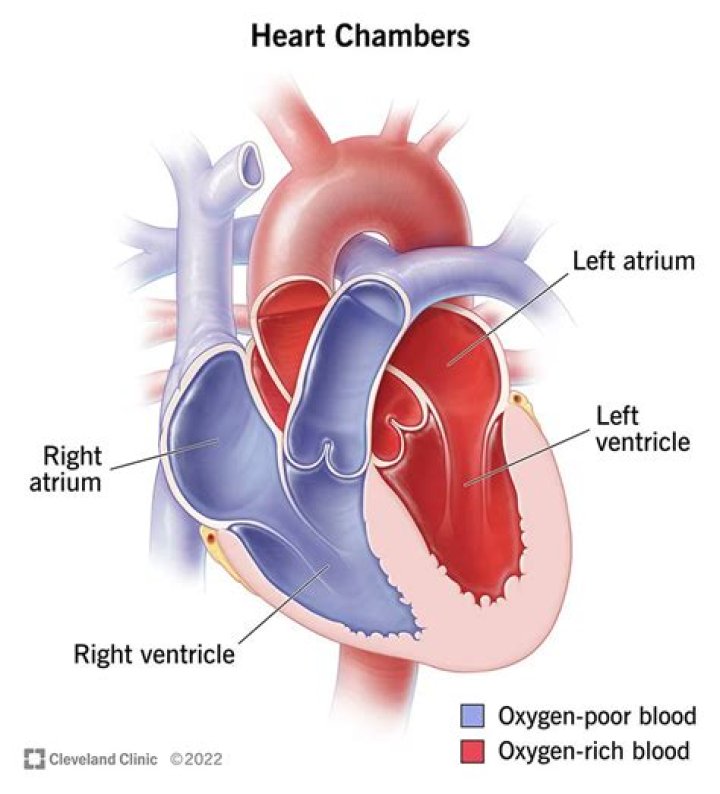
The two atria are thin-walled chambers that receive blood from the veins. The two ventricles are thick-walled chambers that forcefully pump blood out of the heart.
What are the function of ventricles?
Function. During systole, the ventricles contract, pumping blood through the body. During diastole, the ventricles relax and fill with blood again. The left ventricle receives oxygenated blood from the left atrium via the mitral valve and pumps it through the aorta via the aortic valve, into the systemic circulation.
What is ventricles in biology?
ventricle, muscular chamber that pumps blood out of the heart and into the circulatory system. Ventricles occur among some invertebrates. … In humans, the ventricles are the two lower chambers of the heart.
What is atria and atrium?
The upper two heart chambers are called atria. Atria are separated by an interatrial septum into the left atrium and the right atrium. The lower two chambers of the heart are called ventricles. Atria receive blood returning to the heart from the body and ventricles pump blood from the heart to the body.Which valves are situated between the atria and ventricles?
The mitral valve and tricuspid valve are located between the atria (upper heart chambers) and the ventricles (lower heart chambers). The aortic valve and pulmonic valve are located between the ventricles and the major blood vessels leaving the heart.
What is ventricle and atrium?
The heart has four chambers. The upper two chambers are the atria, and the lower two are the ventricles (Figure A). The chambers are separated by a wall of tissue called the septum. Blood is pumped through the chambers, aided by four heart valves. The valves open and close to let the blood flow in only one direction.
Why are the walls of the atria and ventricles different?
The ventricles of the heart have thicker muscular walls than the atria. This is because blood is pumped out of the heart at greater pressure from these chambers compared to the atria. … This is due to the higher forces needed to pump blood through the systemic circuit (around the body) compared to the pulmonary circuit.
Which is the largest artery in the body?
Aorta Anatomy The aorta is the large artery that carries oxygen-rich blood from the left ventricle of the heart to other parts of the body.Why are valves called Semilunar?
The semilunar valves are flaps of endocardium and connective tissue reinforced by fibers which prevent the valves from turning inside out. They are shaped like a half moon, hence the name semilunar (semi-, -lunar).
What are the 4 main heart valves?- tricuspid valve: located between the right atrium and the right ventricle.
- pulmonary valve: located between the right ventricle and the pulmonary artery.
- mitral valve: located between the left atrium and the left ventricle.
- aortic valve: located between the left ventricle and the aorta.
What is the difference between atria and ventricles of the heart?
The atrium supplies the heart and the lungs, whereas the ventricles supply the whole body. The atrium contains deoxygenated blood, whereas the ventricles always contain oxygenated blood. The walls of the atrium are thinner, whereas the walls of the ventricles are thicker.
What is the purpose of the endocardium?
Definition and Function Anatomic function: A tissue covering the inside of the heart, the endocardium keeps the blood flowing through the heart separate from the myocardium, or cardiac muscles. It also lines the valves, which open and close to regulate blood flow through the chambers of the heart.
What is the main difference between your ventricles and your atria Brainpop?
What is the main difference between your ventricles and your atria? Your atria bring blood into the heart; your ventricles pump it out.
Is atrium and atria the same?
Atria and atrium aren’t exactly the same although they refer to the same anatomical structure(s).
What separates atria from ventricles?
The atria are separated from the ventricles by the atrioventricular valves: The tricuspid valve separates the right atrium from the right ventricle. The mitral valve separates the left atrium from the left ventricle.
What separates the two atria?
A wall of muscle called the septum separates the left and right atria and the left and right ventricles.
Where is heart located?
It lies in the front and middle of your chest, behind and slightly to the left of your breastbone. It is a muscle that pumps blood to all parts of your body to provide it with the oxygen and nutrients in needs to function. Your heart has the right and left separated by a wall.
What is the pulmonary flow?
Pulmonary circulation moves blood between the heart and the lungs. It transports deoxygenated blood to the lungs to absorb oxygen and release carbon dioxide. The oxygenated blood then flows back to the heart. Systemic circulation moves blood between the heart and the rest of the body.
Why is it called tricuspid and bicuspid?
Atrioventricular Valves There are two AV valves: Tricuspid valve – located between the right atrium and the right ventricle (right atrioventricular orifice). … It is also known as the bicuspid valve because it has two cusps (anterior and posterior).
What is the largest vein?
The largest vein in the human body is the inferior vena cava, which carries deoxygenated blood from the lower half of the body back up to the heart.
Which is the smallest artery in human body?
- Arteries carry blood away from your heart.
- Veins carry blood back toward your heart.
- Capillaries, the smallest blood vessels, connect arteries and veins.
What is blood capillary?
Capillaries are very tiny blood vessels — so small that a single red blood cell can barely fit through them. They help to connect your arteries and veins in addition to facilitating the exchange of certain elements between your blood and tissues.
What is the largest valve in the heart?
The aortic valve opens the way for oxygen-rich blood to pass from the left ventricle into the aorta, your body’s largest artery.
Which valve is a bicuspid?
A bicuspid aortic valve is an aortic valve that has two flaps (cusps) instead of three. It may cause a narrowed or obstructed aortic valve opening (aortic valve stenosis), making it difficult for the heart to pump blood into the body’s main artery (aorta).
What is deoxygenated blood do?
Blood vesselFunctionHepatic veinCarries deoxygenated blood back to the heart. Carries digested food (glucose and amino acids) from the liver around the body.
What is the difference between endocardium and pericardium?
As nouns the difference between endocardium and pericardium is that endocardium is (anatomy|cardiology) a thin serous membrane that lines the interior of the heart while pericardium is (anatomy|cardiology) a serous membrane that surrounds the heart allowing it to contract.
What is pericardium cavity?
The pericardial cavity is the potential space formed between the two layers of serous pericardium around the heart. … The cavity surrounds the heart and is continuous with it at all but the points of entry and exit of great vessels.
What is the difference between myocardium endocardium and pericardium?
Furthermore, the myocardium is responsible for heart contractions while pericardium is responsible for the protection of the heart. Epicardium, myocardium, and endocardium are the three layers of the wall of the heart while pericardium is the membrane that surrounds the heart.