What is the role of Chondroblasts
Chondroblasts, located in the perichondrium, are cells that play an important role in the development of cartilage. By producing extracellular matrix, chondroblasts create the main component that provides structure and strength to cartilage.
What is the role of chondrocytes during bone elongation?
As in Long Bones, Hypertrophic Chondrocytes in the Mandibular Condyle Undergo Direct Transformation into Bone Cells. The MCC is a site of growth and articulation, combining the growth capability of the limb growth plate with the articular function of articular cartilage.
What is the function of chondrocytes Class 11?
Chondrocytes are the cells making up the cartilage. They are pivotal to synthesize cartilage matrix and sustain the extracellular matrix.
What is the description of chondrocytes?
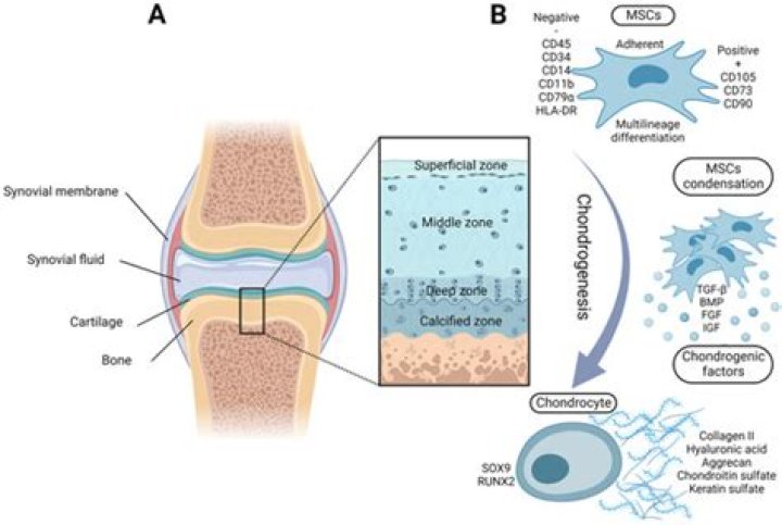
Chondrocytes (/ˈkɒndrəsaɪt, -droʊ-/, from Greek χόνδρος, chondros = cartilage + κύτος, kytos = cell) are the only cells found in healthy cartilage. They produce and maintain the cartilaginous matrix, which consists mainly of collagen and proteoglycans.What is Chondroblast and chondrocytes?
Chondroblasts are a type of cells found in the cartilage which are responsible for the cartilage development. Chondrocytes are a type of specialized cells found in cartilage which are responsible for cartilage maintenance.
What do hypertrophic chondrocytes secrete?
More importantly, hypertrophic chondrocytes secrete critical paracrine factors, such as vascular endothelial growth factor (VEGF) that induce invasion of blood vessels from the perichondrium, and Indian hedgehog (Ihh) that regulate proliferation and differentiation of chondrocytes and directs perichondrial cells to …
What does Chondroblast mean?
[ kŏn′drə-blăst′ ] n. A cell of growing cartilage tissue. chondroplast.
Why do chondrocytes proliferate?
The coordinated proliferation and differentiation of chondrocytes ensures the continuous elongation of the epiphyseal growth plates. The sequential changes between proliferation and differentiation are tightly regulated by secreted growth factors, which activate chondrocyte-specific transcription factors.What is the difference between Osteocyte and chondrocyte?
Osteocytes are developed in the mucoid connective tissue and a mature osteocyte contains a single nucleus. Chondrocytes are involved in the maintenance of cartilage. … Osteocytes are involved in the maintenance of bone tissue. This is the difference between Chondrocytes and Osteocytes.
Is collagen a Fibre?Collagen fiber is the fiber in the extracellular matrix of connective tissues characterized by being elongated and made up of collagen glycoproteins. … It is a strong insoluble fiber. It occurs in the skin, tendon, ligaments, bone, and cartilage.
Article first time published onDo adults have chondrocytes?
As the only cell type found in healthy adult cartilage, chondrocytes are the obvious and most direct starting point for cartilage tissue engineering. Human adult, juvenile, neonatal, and fetal chondrocytes have all been demonstrated to produce cartilage matrix components in vitro for production of engineered tissues.
What is a fibroblast?
fibroblast, the principal active cell of connective tissue. Fibroblasts are large, flat, elongated (spindle-shaped) cells possessing processes extending out from the ends of the cell body.
What are chondrocytes and lacunae?
Chondrocytes, or chondrocytes in lacunae, are cells found in cartilage connective tissue. They are the only cells located in cartilage. They produce and maintain the cartilage matrix, which is a type of lake in which the chondrocytes swim.
What is the shape of chondrocytes?
With a long axis parallel to the cellular surface, juvenile chondrocytes are elliptic in shape at the periphery of cartilages. As the frame shifts inward, the shape takes a round form. Chondrocytes may also appear in isogenous groups of up to eight cells.
How do Chondroblasts become chondrocytes?
Chondroblasts are called chondrocytes when they embed themselves in the cartilage matrix, consisting of proteoglycan and collagen fibers, until they lie in the matrix lacunae.
Does fibrocartilage have Chondroblasts?
Fibrocartilage is a specialized form of connective tissue in which the ground substance is cartilage. … The ground substance between the fibers is basophilic and contains chondroblasts/cytes within lacunae. Fibrocartilage does not contain a perichondrium.
What are Osteoprogenitor cells?
Introduction. Osteoprogenitor cells, also known as osteogenic cells, are stem cells located in the bone that play a prodigal role in bone repair and growth. These cells are the precursors to the more specialized bone cells (osteocytes and osteoblasts) and reside in the bone marrow.
What is the Perichondrium?
Perichondrium is a type of connective tissue, and also functions in the growth and repair of cartilage. Once vascularized, the perichondrium becomes the periosteum. [
What is Chondrogenic cells?
Chondroblasts are progenitor cells that secrete the extracellular matrix (ECM), while chondrocytes are involved in nutrient diffusion and matrix repair. Both cell types are required to form cartilage.
What do osteoblast cells do?
OSTEOBLASTS are the cells that form new bone. … They produce new bone called “osteoid” which is made of bone collagen and other protein. Then they control calcium and mineral deposition. They are found on the surface of the new bone.
What happens to chondrocytes in OA?
Structural Changes in OA Cartilage OA results in progressive cartilage degradation characterized by the softening, fibrillation and erosions of the articular surface [20]. Breakdown of proteoglycans leads to a reduction in the compressive stiffness of the tissue that accelerates the rate of collagen loss [21].
What happens to hypertrophic chondrocytes?
We show that hypertrophic chondrocytes can survive the cartilage-to-bone transition and become osteoblasts and osteocytes during endochondral bone formation and in bone repair.
Why do chondrocytes undergo hypertrophy?
To mediate longitudinal bone growth, round chondrocytes become flattened, proliferate and organize into columns. … These then undergo a tre- mendous increase in cell volume to become hypertrophic chondrocytes (HCs). Mature HCs secrete matrix vesicles, which mediate cartilage calcification.
Where are both chondrocytes and osteocytes located?
Chondrocytes are located in the cartilage of the body and osteocytes are located in the bone.
What is the hypertrophic zone?
The germinal zone of the physis borders the epiphysis. The epiphyseal cartilage cells grow toward the metaphysis and form columns of cells. These columns degenerate, undergo hypertrophy, and then calcify at the metaphysis to form new bone. The hypertrophic zone (shaded red) is the usual site of physeal fractures.
What is the function of compact and spongy bone?
The compact bone is the main structure in the body for support, protection, and movement. Due to the strong nature of compact bone, compared to spongy bone, it is the preferred tissue for strength. Spongy bone is used for more active functions of the bones, including blood cell production and ion exchange.
What happens to the cartilage after it Hypertrophies?
As the inner cartilage hypertrophies and the ossification front extends farther outward, the remaining cartilage in the epiphyseal growth plate proliferates. As long as the epiphyseal growth plates are able to produce chondrocytes, the bone continues to grow.
Do chondrocytes undergo mitosis?
chondrocytes undergo rapid mitotic cell division, enlarge slightly, and become aligned like a stack of coins into longitudinal columns of flattened lacunae. chondrocytes cease dividing and begin to hypertrophy (enlarge) greatly.
Is Endochondral an ossification?
Endochondral ossification is the process by which the embryonic cartilaginous model of most bones contributes to longitudinal growth and is gradually replaced by bone.
What are the 4 types of collagen?
The four main types are type I, II, III, and IV ( 1 ). Here’s a closer look at the four main types of collagen and their roles in your body: Type I. This type accounts for 90% of your body’s collagen and is made of densely packed fibers.
Does coffee reduce collagen?
Caffeine slows down the rate at which your body makes collagen. This is a protein that gives your skin its tightness and elasticity. Once it drops, your skin starts to sag, and wrinkles appear.