What is V5 on a heart monitor
ST segment depression in the left precordial leads, especially V5, can be an indication of myocardial ischaemia, such as can arise during the stress of anaesthesia or operation. As a measure of myocardial oxygen usage, the rate-pressure product was calculated.
Where does V5 lead go?
V5 is placed directly between V4 and V6. V6 is placed over the fifth intercostal space at the mid-axillary line (as if drawing a line down from the armpit). V4-V6 should line up horizontally along the fifth intercostal space.
Why is it called a 12-lead ECG?
The 12-lead ECG displays, as the name implies, 12 leads which are derived by means of 10 electrodes. Three of these leads are easy to understand, since they are simply the result of comparing electrical potentials recorded by two electrodes; one electrode is exploring, while the other is a reference electrode.
What are the leads of ECG?
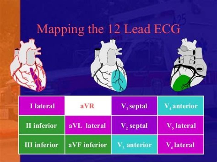
Parts of an ECG The six limb leads are called lead I, II, III, aVL, aVR and aVF. The letter “a” stands for “augmented,” as these leads are calculated as a combination of leads I, II and III. The six precordial leads are called leads V1, V2, V3, V4, V5 and V6. Below is a normal 12-lead ECG tracing.What is V4 V5 V6 in ECG?
The electrical activity on an ECG (EKG). The areas represented on the ECG are summarized below: V1, V2 = RV. V3, V4 = septum. V5, V6 = L side of the heart.
What disorder can an EKG detect?
You may have an EKG as part of a routine exam to screen for heart disease. This test also is used to detect and study heart problems such as heart attacks, arrhythmia or irregular heartbeat, and heart failure. Results from this test also may suggest other heart disorders.
Which ECG machine is best?
ProductPriceProduct BPL 9108D 12 Channel ECG MachinePrice ₹93,000.00Product BPL Cardiart 9108,12 channel ECG MachinePrice ₹99,500.00Product Philips TC20 12 channel ECG MachinePrice ₹158,000.00Product 12 channel ECG Machine , Bionet Model Cardiocare 2000Price ₹60,200.00
What are the three types of ECG leads?
- Limb Leads (Bipolar)
- Augmented Limb Leads (Unipolar)
- Chest Leads (Unipolar)
Where do ECG leads go on chest?
- V1: 4th intercostal space (ICS), RIGHT margin of the sternum.
- V2: 4th ICS along the LEFT margin of the sternum.
- V4: 5th ICS, mid-clavicular line.
- V3: midway between V2 and V4.
- V5: 5th ICS, anterior axillary line (same level as V4)
- V6: 5th ICS, mid-axillary line (same level as V4)
It can record heart activity on six different leads at once (I, II, II, aVL, aVR and aVF). It can detect atrial fibrillation (AFib), bradycardia (abnormally low heart rate) and tachycardia (abnormally high heart rate), but promises to also detect other arrhythmias that could indicate heart disease.
Article first time published onWhat can a 1 lead ECG show?
Although 1-lead ECG (EKG) recorders are normally used primarily for basic heart monitoring, checking for various arrhythmias, or simple educational or research purposes, they can also be used for looking at the effects of exercise on the ECG.
Why do we use lead 2 in ECG?
To assess the cardiac rhythm accurately, a prolonged recording from one lead is used to provide a rhythm strip. Lead II, which usually gives a good view of the P wave, is most commonly used to record the rhythm strip.
Which wave of an ECG will show abnormality in atrial fibrillation?
Diagnosis – Atrial Fibrillation. The diagnosis of atrial fibrillation is confirmed with a standard 12-lead ECG. P waves are absent, coarse “fibrillatory waves” can frequently be seen and sometimes no atrial activity can be identified. The QRS complexes are “irregularly irregular”, with varying R-R intervals.
Where do you place a 5 lead cardiac monitor?
Five-lead system Place the left arm (LA) electrode near the left shoulder, close to the junction of the left arm and torso. Place the right leg (RL) electrode below the level of the lowest rib on the right abdominal area. Place the left leg (LL) electrode below the level of the lowest rib on the left abdominal area.
Are EKG and ECG the same?
An electrocardiogram records the electrical signals in your heart. It’s a common and painless test used to quickly detect heart problems and monitor your heart’s health. Electrocardiograms — also called ECGs or EKGs — are often done in a doctor’s office, a clinic or a hospital room.
Which lead is most important to record in a 1 year old?
For infants, toddlers, and children under 90 lbs, measuring rib spaces is not usually possible. For all ECGs, limb leads should be placed on the limbs — not the torso. Arm leads should be placed just above the elbows. Leg leads should be placed between the knee and ankle.
What does ischemia look like on an ECG?
The most common ECG sign of myocardial ischemia is flat or down-sloping ST-segment depression of 1.0 mm or greater. This report draws attention to other much less common, but possibly equally important, ECG manifestations of myocardial ischemia.
Are V1 V6 unipolar or bipolar?
The electrode leads each have a name. The bipolar extremity leads are called I, II and III. The unipolar extremity leads are called avR, avL and avF, and the chest leads are called V1–V6.
Why does hypertension increase left ventricular size?
Your heart muscle cells may get larger in response to some factor that causes the left ventricle to work harder, such as high blood pressure or a heart condition. As the left ventricle’s workload increases, the muscle tissue in the chamber wall thickens. Sometimes, the size of the chamber itself also increases.
Can I do an ECG at home?
Having a patient being able to take an electrocardiogram at home can be very helpful and important as part of a cardiologist’s evaluation via telemedicine. The two most common and practical ways for a patient to do an EKG on themselves are via an app called Kardia or with a later series (4 or 5) Apple Watch.
Can ECG be done at home?
What is [email protected]? Electro encephalogram or ECG is test using a specialized machine. This test measures the heart beat and monitors the activities of a person’s heart. Usually these machines are found at diagnostic centres and hospitals, but now, small portable variations can also be used at home.
Can we do ECG test at home?
A home or personal-use ECG can be a helpful tool if you have a condition that might affect your heart rate and rhythm. Let your doctor know if you have any unusual ECG readings. A personal-use ECG monitor won’t be as accurate as a clinical or hospital-grade ECG machine.
Can an ECG detect a blocked artery?
An ECG Can Recognize the Signs of Blocked Arteries. Unfortunately, the accuracy of diagnosing blocked arteries further from the heart when using an ECG decrease, so your cardiologist may recommend an ultrasound, which is a non-invasive test, like a carotid ultrasound, to check for blockages in the extremities or neck.
Can EKG detect blocked arteries?
No, an electrocardiogram cannot detect blocked arteries. Blocked arteries are usually diagnosed with a nuclear stress test, cardiac pet scan, coronary CT angiogram or traditional coronary angiogram.
What does an ECG tell you about your heart?
An ECG (electrocardiogram) records the electrical activity of your heart at rest. It provides information about your heart rate and rhythm, and shows if there is enlargement of the heart due to high blood pressure (hypertension) or evidence of a previous heart attack (myocardial infarction).
How often should ECG leads be changed?
Electrodes should be changed daily. Electrode placement is integral for accurate results. When an electrode is misplaced by as little as one intercostal space, QRS morphology may change and contribute to misdiagnosis.
How do you perform a female ECG?
Small pads or patches (electrodes) will be placed, like stickers, on your skin on each arm and leg and on your chest. The electrodes are hooked to a machine that traces your heart activity onto a paper. During the test, lie very still and breathe normally. Do not talk during the test.
Why is aVR inverted?
The aVR is often neglected lead. It is an unipolar lead facing the right superior surface. As all the depolarisations are going away from lead aVR, all waves are negative in aVR (P, QRS, T) in normal sinus rhythm.
When is a 3 lead ECG used?
3-lead ECGs are used most often for recording a 24-hour reading. A 24-hour reading is a frequently used tool for the diagnosis of heart problems and is reimbursed as a long-term reading.
What are the 12 leads of ECG?
The standard EKG leads are denoted as lead I, II, III, aVF, aVR, aVL, V1, V2, V3, V4, V5, V6. Leads I, II, III, aVR, aVL, aVF are denoted the limb leads while the V1, V2, V3, V4, V5, and V6 are precordial leads.
How does a KardiaMobile 6L work?
To use the KardiaMobile, you simply place your fingers on the two device sensors for 30 seconds. No electrodes are needed. The results are immediately compiled and then displayed on your smartphone. From there, you have the option to email these results to your doctor.