Where is the greater Petrosal
The greater petrosal nerve (or greater superficial petrosal nerve) is a nerve in the skull that branches from the facial nerve; it forms part of a chain of nerves that innervate the lacrimal gland. The preganglionic parasympathetic axons of this nerve synapse in the pterygopalatine ganglion.
What does greater petrosal nerve do?
The greater petrosal nerve or superficial petrosal nerve is a branch of the nervus intermedius (nerve of Wrisberg) that carries parasympathetic, taste, and sensory fibers of the facial cranial nerve (CN VII).
Does the greater petrosal nerve go through the foramen Lacerum?
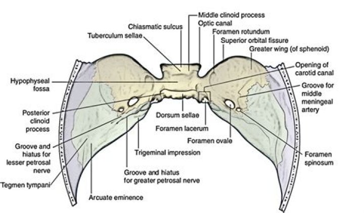
In the middle cranial fossa, the greater petrosal nerve passes medially to enter the foramen lacerum and fuses there with the deep petrosal nerve, forming the Vidian nerve or pterygoid nerve, which passes from the pterygoid canal to the pterygopalatine fossa (PPF).
Where does the greater petrosal nerve synapse?
The greater petrosal nerve exits the canal with the deep petrosal nerve and synapses in the pterygopalatine ganglion in the pterygopalatine fossa. The ganglion then gives of nerve branches which supply the lacrimal gland and the mucous secreting glands of the nasal and oral cavities.Where does the deep petrosal nerve come from?
The deep petrosal nerve is a branch from the internal carotid plexus. The plexus is located on the lateral side of the internal carotid as it courses superiorly. The deep petrosal enters the skull through the carotid canal with the internal carotid artery.
What is the hypoglossal?
The hypoglossal nerve enables tongue movement. It controls the hyoglossus, intrinsic, genioglossus and styloglossus muscles. These muscles help you speak, swallow and move substances around in your mouth.
Where does the lesser petrosal nerve come from?
The lesser petrosal nerve (Figure 26.3) is a continuation of the presynaptic fibers of the tympanic branch of the glossopharyngeal nerve with contributions from the nervus intermedius part of the facial nerve, and the auricular branch (Alderman’s or Arnold’s nerve) of the vagus nerve.
What is the ciliary ganglion?
Ciliary ganglion is a peripheral parasympathetic ganglion. It is situated near the apex of orbit between the optic nerve and lateral rectus muscle. It is related medially to the ophthalmic artery and laterally to the lateral rectus muscle.Where is the chorda tympani located?
The chorda tympani is a nerve that arises from the mastoid segment of the facial nerve, carrying afferent special sensation from the anterior two-thirds of the tongue via the lingual nerve, as well as efferent parasympathetic secretomotor innervation to the submandibular and sublingual glands.
Where does the facial nerve enter the cranium?It emerges from the pons of the brainstem, controls the muscles of facial expression, and functions in the conveyance of taste sensations from the anterior two-thirds of the tongue. The nerves typically travels from the pons through the facial canal in the temporal bone and exits the skull at the stylomastoid foramen.
Article first time published onWhat is the tympanic nerve?
Medical Definition of tympanic nerve : a branch of the glossopharyngeal nerve arising from the petrosal ganglion and entering the middle ear where it takes part in forming the tympanic plexus. — called also Jacobson’s nerve.
Where is the tympanic plexus?
In the middle ear, the tympanic plexus is formed on the tympanic promontory by branches of Jacobson’s nerve (tympanic branch of the glossopharyngeal nerve) and caroticotympanic nerves originating from the internal carotid artery plexus.
What nerves pass through the foramen ovale?
The foramen ovale transmits the mandibular nerve, accessory meningeal artery, lesser petrosal nerve and the emissary veins.
What passes through hypoglossal canal?
Function. The hypoglossal canal transmits the hypoglossal nerve from its point of entry near the medulla oblongata to its exit from the base of the skull near the jugular foramen.
What passes through the foramen cecum?
(Foramen cecum is third label from the top.) … The foramen cecum varies in size in different subjects, and is frequently impervious; when open, it transmits the emissary vein from the nose to the superior sagittal sinus.
Is the deep petrosal nerve in the middle cranial fossa?
Leaving the geniculate ganglion, the greater petrosal nerve pierces the upper surface of petrous temporal bone, enters the middle cranial fossa, is joined at foramen lacerum by the deep petrosal nerve (sympathetic fibres from internal carotid plexus) to form the nerve of the pterygoid canal, which goes to the …
What is external petrosal nerve?
The external petrosal nerve is one of the three branches from the geniculate ganglion. It carries sympathetic fibers to the sympathetic plexus surrounding the middle meningeal artery, coursing extradurally laterally to the greater and lesser petrosal nerves on the petrous ridge’s anterior surface 3.
What is the lesser petrosal n?
The lesser petrosal nerve (also known as the small superficial petrosal nerve) is the general visceral efferent (GVE) component of the glossopharyngeal nerve (CN IX), carrying parasympathetic preganglionic fibers from the tympanic plexus to the parotid gland.
Where do the preganglionic parasympathetic fibers of lesser petrosal nerve switch on postganglionic fibers?
The preganglionic parasympathetic fibers synapse with the cell bodies of the postganglionic parasympathetic fibers in the otic ganglion located on the V3 (medial side), around the origin of the nerve to the medial pterygoid (10).
What is Jacobson nerve?
Jacobson’s nerve is a tympanic branch of the glossopharyngeal nerve, arising from its inferior ganglion. It enters the middle ear cavity through the inferior tympanic canaliculus, runs in a canal on the cochlear promontory and provides the main sensory innervation to the mucosa of the mesotympanum and Eustachian tube.
What does the Abducens nerve do?
The abducens nerve functions to innervate the ipsilateral lateral rectus muscle and partially innervate the contralateral medial rectus muscle (at the level of the nucleus – via the medial longitudinal fasciculus).
Is hypoglossal nerve a mixed nerve?
CN XII, Hypoglossal, innervates the muscles of the throat and enables us to swallow. Five cranial nerves have mixed sensory, motor and parasympathetic function.
Are there nerves in the tongue?
The tongue has many nerves that help detect and transmit taste signals to the brain. Because of this, all parts of the tongue can detect these four common tastes; the commonly described “taste map” of the tongue doesn’t really exist.
What is the origin of the chorda tympani nerve?
The chorda tympani nerve arises from the facial nerve a few millimeters above the stylomastoid foramen. It is directed superior and anterior, and perforates the tympanic cavity. It enters the posterior canaliculus and then descends near the spine of the sphenoid bone.
Where does the lingual nerve come from?
The lingual nerve carries sensory innervation from the anterior two-thirds of the tongue. It contains fibres from both the mandibular division of the trigeminal nerve (CN V3) and from the facial nerve (CN VII).
Why is tympani called chorda?
Structure and Location. After splitting off from the intracranial branch of the facial nerve, the chorda tympani enters the ear. Its association with the ear is what gives the chorda tympani its name.
What is ophthalmic nerve?
The ophthalmic nerve is the first branch of the trigeminal nerve, which is also known as the fifth cranial nerve. The ophthalmic nerve supplies sensory innervation to the structures of the eye, including the cornea, ciliary body, lacrimal gland, and conjunctiva.
Where is ciliary muscle?
The ciliary muscle is elongated, triangular in shape, and located beneath the anterior sclera just posterior to the limbus. The shortest side of the triangular region faces anterior-inward and it is to this region of the ciliary body that the base of the iris inserts.
What are the 4 parasympathetic ganglia?
Location of Autonomic Ganglia Parasympathetic ganglia which innervate targets in the head are located in four main ganglia: the ciliary, pterygopalatine, submandibular and otic ganglia. Scattered microganglia may also be distributed along cranial nerves.
Where are the nerves in your face located?
The facial nerve exits the base of the skull at the stylomastoid foramen, which is an opening in the bone located near the base of the ear.
Where is the 7th facial nerve located?
The two 7th Cranial Nerves (CN VII) are located on either side of the brainstem, at the top of the medulla. They are mixed cranial nerves with BOTH sensory and motor function. CN VII controls the face and is mainly FACE MOVEMENT with some face sensation.